
The gastrocnemius, soleus and plantar muscles form the triceps muscle of the calf, which transforms into the Achilles tendon that attaches to the calcaneus. The function of the Achilles tendon is to transfer the kickback force from the calf muscles to the foot while walking and running. Achilles tendon pathologies are most often the result of overloading the tendon or undertaking intensive training after a long break without proper preparation. This leads to unfavorable changes in the structure of the tendon or inflammation of its sheath. Failure to treat the Achilles tendon properly may result in rupture of the tendon. The complete disruption of the tendon is treated by suturing with a minimally invasive method or with the open method.
Make an appointment now - with a specialist in Achilles tendon pain management at our hospital
[title]
[image-intro]
[readmore text="Read more"]{/article}
[title]
[image-intro]
[readmore text="Read more"]{/article}
[title]
[image-intro]
[readmore text="Read more"]{/article}
[title]
[image-intro]
[readmore text="Read more"]{/article}
Achilles tendon diseases
Achilles diseases belong to the groups of the so-called tendinopathy, i.e. pathological conditions affecting the muscle tendons. Achilles tendinopathy is the result of the aggregation of microdamages that occur when the tendon is overloaded. Abnormal lesions usually cover the 2-6 cm section of the tendon above the heel bone attachment to the tumor, which is associated with poor blood supply to this area. A factor that further disturbs the blood supply to the tendon is excessive valgus of the hindfoot manifested by too much leaning of the heels inward. In people with this foot defect, the Achilles tendon twists undesirably when walking or running, causing the blood vessels to close. Another risk factor for tendinopathy is increased calf triceps tension and Achilles contracture resulting from too short recovery time and neglect of stretching exercises.
Achilles tendon degeneration (tendinosis)
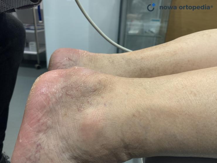
Achilles tendinosis is the degeneration of the interior of the tendon without signs of inflammation. Repeated damage to the tendon tissue leads to local foci of necrosis and disturbance of the fibril structure of the tendon, as a result of which its mechanical strength is reduced. On a clinical examination, you can feel a thickened section of the tendon as it moves through the flexion and extension movements of the foot. Tendon palpation and Achilles pain when walking or running are present. The ultrasound examination shows the disorganization of the fiber path, the ingrowth of new blood vessels and calcification centers. In MRI on T1-weighted images, tendinosis changes are visible as irregular focal amplification of the signal within the tendon with increased circumference. In some cases, "tendinosis" may present as Achilles tendon enthesopathy - a degenerative condition at the Achilles tendon attachment to the calcaneus. This may be accompanied by an inflammation of the bursa. Tendon pathology is more common in the presence of the so-called Haglund's heel, i.e. bone growth located in the upper-lateral part of the Achilles attachment. Bone outgrowth can mechanically irritate soft tissues and thus contribute to the development of tendinosis.
Tendinosis and Achilles tendinitis
The term "tendinitis" for "Achilles inflammation" was overused until recently. Some authors use the term "tendinitis" to mean the state of exacerbation of tendinosis, which is manifested by a greater degree of microdamage to the tendon and more intense pain. However, it is now known that inflammation does not occur in the course of tendinosis. "Tendinitis" is a rare pathology in which the Achilles tendon is actually inflamed - its diagnosis and differentiation from tendinosis is very important due to different therapeutic procedures.
Insertional tendinopathy
Achilles tendon longitudinal section
Achilles tendon cross-section
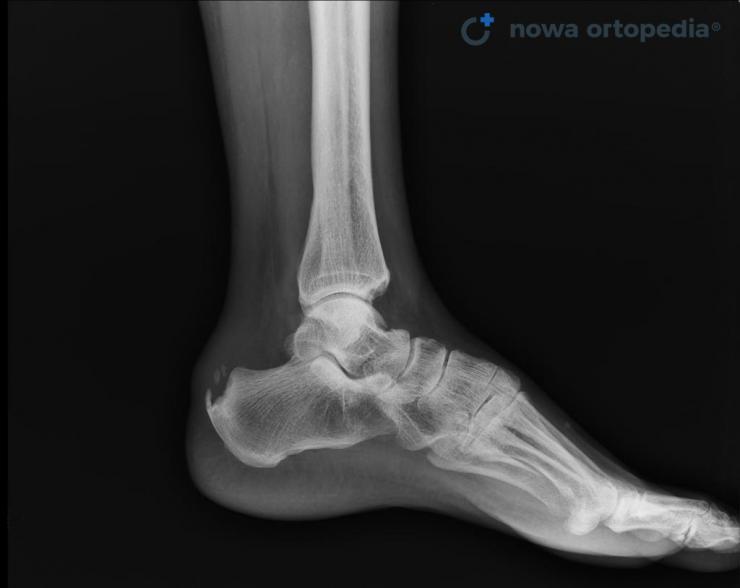
X-ray image - calcification in Achilles
Inflammation of the tendon sheath (paratenonitis)
The Achilles tendon has a sheath called parathenone, which allows the tendon to slide freely inside it. Paratenone also extends to 2-3 cm with the elongation of the Achilles tendon. Symptoms of sheath inflammation are local pressure soreness and fluid accumulation in the tendon area. There is local pain in the Achilles tendon with foot movements, starting in the morning and increasing with activity. Acute Achilles' tenosynovitis lasts up to about 2 weeks, when the symptoms are most severe, then the symptoms are milder and may recur with less intensity, e.g. at the beginning of the run. After inflammation, the sheath is scarred, it may "stick" to the Achilles tendon, disrupting its mutual displacement.
Treatment of Achilles tendon diseases
The first step in improving the condition of the Achilles tendon is to reduce training loads, e.g. reducing the weekly running mileage. The aim of conservative treatment of tendinopathy is to improve the flexibility, blood supply and metabolism of the Achilles tendon, as well as to stimulate the processes of reconstruction of the tendon fibers. A modern method of treating Achilles tendinopathy is the Topaz coblation procedure, which uses radio waves to trigger a controlled inflammatory reaction and stimulate the proper healing and repair processes of tissues.
Manual therapy conducted as part of rehabilitation usually includes deep transverse massage of the tendon and functional massage of the rear part of the calf. Other fascial techniques that affect the entire posterior superficial tape can also be used to normalize the muscle tone of the posterior shank group. The effect of manual work should be preserved by using kinesiotaping, i.e. tapes glued on the skin. As part of home self-therapy, the patient should perform exercises to stretch the calf muscles and the so-called eccentric training according to Alfredson. This training involves the controlled lowering of the heel down below the level of the step on which the forefoot rests. The patient performs 3 series of 15 repetitions in the morning and evening for a minimum period of 3-6 months.
As part of physical therapy, the treatment of tendinosis includes, among others, shock wave therapy, ultrasound and cryotherapy. In the case of valgus heels, individual orthopedic insoles should be obtained, which will improve the biomechanics of the ankle joint and reduce the impact of torsional forces on the Achilles tendon.
Injecting steroids into the tendon is inadvisable as they may widen the necrosis and lead to Achilles tendon rupture.
Surgical treatment is rarely performed and consists in the removal of necrotic tissues within the Achilles tendon - often created as a complication of steroid administration.
Sources:
1. Bass E. Tendinopathy: Why the Difference Between Tendinitis and Tendinosis Matters. International Journal of Therapeutic Massage & Bodywork. 2012;5(1):14-17.
Frequently asked questions about Achilles tendon pain:
In the case of Achilles tendon pain, an orthopedist should be consulted, who will accurately diagnose the cause of the ailments and establish a treatment plan. The management consists mainly of a temporary limitation of sports activity, performing eccentric calf muscle exercises and procedures in the field of physical therapy. In the treatment of chronic Achilles tendinopathy, modern methods are used to stimulate the processes of proper healing and tissue repair, e.g. the Topaz electrode coblation procedure.
Strengthening the Achilles tendon by stimulating the reconstruction of the fiber structure can be achieved by performing eccentric training according to the Alfredson protocol. This training involves the controlled lowering of the heel down below the level of the step on which the forefoot rests. The exercise is performed with the knee extended and then bent. The training includes 15 repetitions in 3 series in the morning and evening for a minimum period of 3-6 months. To be sure that the exercise is being performed correctly, it's best to check with your physical therapist.


