
Pressure syndromes, also known as pressure neuropathies, are pathological conditions involving disturbance of the nerve's function as a result of mechanical irritation. Nerve compression can be caused by increased muscle mass, post-traumatic edema, inflammation, connective tissue scar or degenerative changes. The conflict can get worse in certain body positions and with certain activities.
The symptoms of pressure are tingling, numbness or pain in the area supplied by the pinched nerve (the most frequently reported symptoms are numbness of the fingers). When tissue conflict leads to long-term nerve conduction disturbances, specific muscles weaken and gradually decline. Nerve entrapment sites are usually the wrist and elbow, where the nerves run in narrow spaces bounded by tissues that are often overloaded or damaged. Conservative treatment of neuropathy remains ineffective in most cases, so management is mainly based on surgical release of the nerve by cutting or removing the tissue causing compression.
Make an appointment now - to a doctor specializing in upper limb nerve decompression at our hospital
[title]
[image-intro]
[readmore text="Read more"]{/article}
[title]
[image-intro]
[readmore text="Read more"]{/article}
[title]
[image-intro]
[readmore text="Read more"]{/article}
Compression of the ulnar nerve
Symptoms of ulnar neuropathy are sensory disturbances in the 4th and 5th finger and the outer part of the hand and forearm. Muscle weakness affects the ball of the little finger, the interosseous muscles, some of the ascaris muscles, and some of the muscles of the thumb ball. As a result, the stability of the fingers during gripping is impaired and it is difficult to handle the objects.
Guyon's Channel Syndrome
The ulnar nerve at the wrist runs in the so-called Guyon's canal, which is bounded by the pea-like bone and hook bone of the wrist, and the flexor cord. Guyon's canal syndrome usually develops as a result of the chronic leaning of the outer wrist area on a grip or a bar.
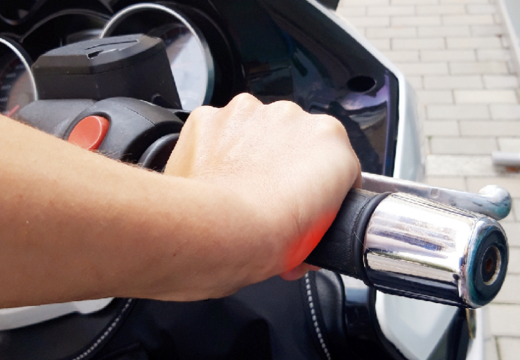
So, neuropathy often occurs in people who walk on crutches, weightlifters and cyclists. Nerve compression may also occur due to inflammatory changes (rheumatoid hand) or degenerative changes, when bone spurs come into conflict with the ulnar nerve. The presence of nodules (ganglions, lipomas) can also compress the nerve and trigger ailments. On the other hand, the sudden appearance of pressure symptoms results from an acute trauma, e.g. bone fracture with dislocation of the fragments or the formation of a traumatic hematoma limiting the space in the Guyon's canal.
Ulnar nerve groove syndrome
The ulnar nerve groove is located on the posterior surface of the medial epicondyle of the humerus (medial side of the elbow). The ulnar nerve is located in a narrow bone groove and covered with a medial collateral ligament. The ulnar nerve groove syndrome is common among players who perform javelin throws or those who practice shot put. Unnaturally increased tension in the ulnar nerve occurs during forced elbow valgus or an elbow flexion swing prior to the roll. Then the ulnar nerve may move out of the groove, return it to its place and thus mechanical irritation of the nerve. Elbow injuries leading to damage and scarring of the elbow ligaments and secondary degenerative changes in the elbow joint predispose to nerve entrapment under connective tissue bands or to nerve compression by bone growth (osteophyte). In addition, pitchers often overload the wrist flexor muscles attached to the medial epicondyle of the humerus. Degenerative changes in the tendon attachments of the muscles may additionally narrow the space around which the ulnar nerve runs. The ulnar nerve groove syndrome is an occupational disease - similar changes as in the case of athletes may occur in workers who perform work involving the upper limbs. In addition, ulnar neuropathy is found in people who habitually keep their elbow constantly bent and rest it on the table top (office workers).
Traumatic fractures of the distal end of the humerus in the medial epicondyle may also cause pressure on the nerve and even lead to a break in its continuity.
Ulnar nerve groove syndrome
Compression of the median nerve
Symptoms of compression of the median nerve are numbness, burning, and pain in the palmar side of the thumb, the second and third fingers, and the middle of the fourth finger on the thumb side. Nerve entrapment can occur in the carpal tunnel or between the muscles at the top of the forearm.
Carpal tunnel syndrome
The carpal tunnel is a bone-fibrous canal limited at the bottom by a series of bones, and at the top by the transverse ligament of the wrist. The median nerve and tendons of the muscles flexing the fingers run in the canal. The presence of inflammation in the course of tenosynovitis, post-traumatic edema or fibrosis of the transverse ligament of the wrist causes a narrowing of the space in which the median nerve runs. The carpal tunnel also develops as a result of frequent resting the hand on the crutch holder, performing manual work or everyday work on the computer.
Read more about carpal tunnel syndrome.
Traumatic fractures of the distal end of the humerus in the medial epicondyle may also cause pressure on the nerve and even lead to a break in its continuity.
The pronator syndrome
The second cause of pressure on the median nerve is its conflict with the muscles or fascia in the upper part of the forearm. To initially diagnose this level of neuropathy, provocation tests are performed:
resisting forearm conversion (the palmar side of the hand goes to the floor) with simultaneous extension of the elbow - nerve entrapment between the two heads of the inverted muscle,
resistance tension of the biceps (biceps muscle of the shoulder) when flexing and inverting the forearm (the palmar side of the hand goes to the ceiling) - compression of the median nerve under the fibrous cord constituting the extension of the biceps tendon,
third finger resistance flexion - entrapment of the median nerve by the arch of the superficial flexor muscle of the fingers.
Compression of the radial nerve
The compression of the radial nerve occurs most often during sleep, when the upper limb is crushed by the weight of your own body. Radial nerve compression is also called "Saturday Night Palsy" because it often happens after alcohol abuse and falling asleep in a random upper limb position. A typical symptom of compression of the radial nerve is the so-called "Drooping hand" because the muscles that straighten the wrist are paralyzed or paralyzed. In the case of this type of neuropathy, the symptoms are usually transient - manual therapy of soft tissues and specific movements in the joints of the upper limb to neuromobilize the nerve help in restoring the hand function.
Nerve decompression surgery
Eligibility for Nerve Release Surgery
Guyon's canal syndrome, ulnar nerve groove syndrome, carpal tunnel syndrome or pronator syndrome are initially diagnosed on the basis of characteristic symptoms - sensory disturbances in a specific area and possible weakening of muscle strength. The diagnosis is confirmed by a nerve conduction test (electrophysiological examination). To determine the cause of pressure on a nerve, the required diagnostic method is soft tissue ultrasound, magnetic resonance imaging (soft and bone tissue), or X-ray (bone tissue only). If there is a conflict between the nerve and the surrounding tissue, a decision is made to decompress the nerve surgically.
The course of the nerve decompression procedure
Nerve release surgery is usually performed using the classic open method, which requires a 2-3 cm incision on the palmar side of the wrist or a few centimeters incision along the nerve path around the elbow. Incision of the skin allows for an accurate assessment of the nerve area and precise excision of conflict-causing tissues. In the endoscopic method, in which surgical instruments are inserted through small holes, the scope of the operating field is limited. Therefore, the endoscopic procedure is performed only in the case of simple procedures involving the cutting of shrunken tissues colliding with the nerve. Most of the procedures that require tissue resection and careful preparation of the space around the nerve are performed using the open method. During the procedure, ligaments are cut, e.g. the transverse ligament of the wrist, tissue necrotic foci and fragments of bones pressing the nerve are removed. The wound is closed with sutures, which are removed 10-14 days after the procedure.
After treatment
Home discharge usually takes place on the day of surgery. The patient receives recommendations from the physiotherapist regarding the care and position of the operated limb. Until the sutures are removed, limb movements should be performed within a limited painless range. After removing the stitches, it is very important to mobilize the postoperative scar in order to avoid connective tissue adhesions and obtain a good cosmetic effect (reducing the visibility of the scar). The patient should perform exercises to support the shifting of the nerve (neuromobilization) - initially, the exercises are carried out under the supervision of a physiotherapist on an outpatient basis, and then continued by the patient at home. Overload of the operated limb should be avoided for several weeks after the surgery. It is advisable to perform safe exercises recommended by a physiotherapist, which are designed to gradually restore the mobility of the elbow and wrist joints and restore the arm's muscular strength. Only when the function of the hand is restored, can you return to professional work or sports.
In the case of partial damage to the nerve structure, it is recommended to take drugs that support the metabolism of the nervous tissue, e.g. preparations rich in B vitamins.
Important information
| Duration of the procedure (depending on the method) | 15 -75 minutes |
| Tests required for surgery | basic or not - depending on the type of anesthesia |
| Anesthesia | local or perimeter block, sectional |
| Hospital stay | up to 1-6 hours after the procedure |
| A period of significant dysfunction | up to 2 weeks |
| A period of limited dysfunction | 2 - 6 weeks |
| Removal of stitches - first visit | 12 - 16 days or none |
| Change of dressings | every 3 - 4 days |
| Contraindications to the procedure | infection |
Frequently asked questions about compression neuropathies and nerve decompression surgery:
Finger numbness is usually a symptom of neuropathy, i.e. dysfunction of the peripheral nerves. Numbness of individual fingers in one hand is most often the result of pressure on the nerve by other tissues, e.g. inflammatory edema, connective tissue scars or degenerative changes. Within the upper limb, places of frequent nerve compression are the wrist and the elbow joint area. The cause of sensory disturbances may also be compression of the nerve root due to degenerative changes in the cervical spine. Symmetrical numbness of the fingers, on the other hand, may indicate a systemic cause of neuropathy, e.g. diabetes, which leads to the weakening of nerve conduction in all the smallest nerves of the human body. In the event of the appearance of the above-mentioned symptoms, it is necessary to consult a doctor who will conduct a detailed diagnosis and establish a treatment plan.
The surgical procedure in the course of the ulnar nerve groove consists in removing the cause of the nerve compression. It is usually a resection of degenerative bone lesions or connective tissue scars limiting the displacement of the ulnar nerve. The procedure requires a few centimeters incision along the nerve path in order to carefully work out the area of the medial area of the elbow. After the surgery, rehabilitation is necessary, which helps to restore the proper slide of the nerve and allows the weakened muscles of the upper limb to be strengthened.
When local inflammation is the cause of nerve compression, therapy is aimed at reducing the inflammation and rapidly evacuating the edema. It is advisable to take anti-inflammatory drugs and to temporarily relieve the limb. However, when post-inflammatory tissue adhesions have occurred or the cause is a bone change or a growing tumor, surgical removal of the lesion is necessary. In the case of overload neuropathies, it is recommended to rest and give up the activity that aggravates the symptoms. In patients who have relapsed neuropathy, surgery is the only effective method of permanent cure.


