
Ultrasound examination of the ankle joint enables the assessment of selected congenital defects, as well as many changes on the basis of overload and inflammation. The ankle ultrasound also allows for the diagnosis of damage to the soft tissues surrounding the joint - ligaments, tendons, muscles, nerves and vessels, as well as the assessment of bone contours and cartilage to a limited extent. It is recommended that ultrasound of the ankle is performed after any torsion of the ankle. Due to the large number of structures in this area, ultrasound should be directed by a clinical examination to quickly make an appropriate diagnosis, and in case of doubt, order an MRI.
Make an appointment now - to the doctor who performs ultrasound of the ankle joint in our hospital
[title]
[image-intro]
[readmore text="Read more"]{/article}
[title]
[image-intro]
[readmore text="Read more"]{/article}
[title]
[image-intro]
[readmore text="Read more"]{/article}
[title]
[image-intro]
[readmore text="Read more"]{/article}
What diseases and injuries can be diagnosed by ultrasound of the ankle joint?
The following are the most common pathologies that can be partially or fully assessed during an ultrasound examination of the ankle joint.
Pain in the front of the ankle:
- Anterior wedging syndrome - the presence of osteophytes limiting the dorsiflexion of the ankle joint (anterior conflict),
- Injury of the anterior tibial tendon,
Pain in the side of the ankle:
- Damage to the lateral ligaments of the ankle joint due to ankle sprain,
- peroneal tendinopathy,
- tendon sheath inflammation of the peroneal tendons
- subluxation of tendons of the fibula muscles,
Medial Ankle Pain:
- damage to the triangular ligament,
- injuries of the posterior tibial tendon,
- tarsal canal syndrome - compression of the posterior tibial nerve - symptoms may also be present in the sole of the foot,
Pain in the back of the ankle joint:
- The presence of an additional triangular ankle (to a limited extent),
- Impaction syndrome - compression of the soft tissues with plantar flexion of the foot,
- Fracture of the lateral process of the talus (snowboarder's fracture),
- Achilles tendinopathy,
- Achilles tendon rupture,
- Achilles tendon bursitis,
Pain in the middle of the ankle joint or diffuse joint pain:
- Synovitis,
- Free bodies at the pond.
The ultrasound of the ankle joint cannot assess:
- tibia cartilage, a block of the talus in 2/3 of the posterior part, and cartilage of the fibula (this refers to the cartilage of the ankle joint),
- intraosseous changes, subchondral changes (bone marrow edema),
- pathology and damage within the tarsal sinus,
- the subtalar joint.
How is ultrasound examination of the ankle joint?
Standard ultrasound examination of the ankle joint includes the assessment of the following areas of the joint: anterior, lateral, medial and posterior. During the examination, the patient lies on his back with the limb bent at the knee to an angle of 45 degrees. During ultrasound of the Achilles tendon and the plantar side of the foot, the patient lies on his stomach.
Dynamic ultrasound examination of the ankle joint is based on the assessment of selected structures during stress tests, joint movement or muscle tension. The capacity of the ligaments is most often assessed, e.g.
- during the anterior drawer test, the anterior talo-fibulare ligament (ATFL) is assessed - most often damaged as a result of ankle sprain,
- during dorsiflexion combined with external rotation of the foot, the inferior anterior tibiofibular ligament (AITFL) and tibiofibular ligament are assessed.
A dynamic examination of the tendons may, for example, concern the tendons of the peroneal muscles - in the case of their habitual subluxation, ultrasound shows abnormal displacement during the flexion and conversion of the foot.
Sonosurgery
Sonosurgery is the term for a percutaneous intervention performed under ultrasound guidance.
Surgery of the ankle joint
Examples of sonosurgery in the ankle area are:
- Puncture of the ankle or ankle joint to collect the synovial fluid,
- Intra-articular blockade - administration of steroids to the joint to reduce pain,
- Administration of steroids into the tendon sheaths lying in the tarsal canal (treatment of inflammation of the sheaths),
- Administration of the steroid to the inflamed bursa of the Achilles tendon (steroid injection of the Achilles tendon itself is contraindicated due to the weakening of the tendon structure and an increased risk of rupture in the future).
Frequently asked questions about ultrasound of the ankle joint:
In order to exclude an ankle fracture, an X-ray of the ankle joint is recommended. This is especially recommended in the case of severe pain and massive swelling and the inability to load the limb in the three-step test. Ankle ultrasound should be performed in every case of ankle sprain to assess the efficiency of the damaged ligaments and to exclude the presence of complications in the pathology of other soft tissues.
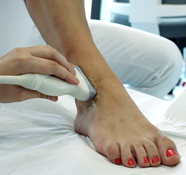
Ankle ultrasound examination is a non-invasive imaging examination using ultrasound waves. During the ultrasound examination, the radiologist or orthopedist applies a special head to the ankle, first applying a coupling gel to the skin of the examined area. All soft tissues surrounding the ankle (ligaments, muscle tendons, straps, nerves, blood vessels) are assessed. Ultrasound images also show the synovium and the contours of the anterior part of the Talus. During the so-called dynamic ultrasound examination, the doctor performs special tests to assess selected structures (e.g. the efficiency of ligaments). It may also ask you to tighten a muscle or make a specific movement with your foot.


