
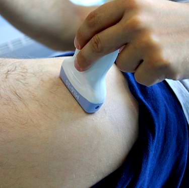
Adult hip ultrasound is a readily available non-invasive diagnostic method that allows the diagnosis of major hip pathologies. Hip ultrasound is useful e.g. in confirming inflammation, the presence of joint exudate and abnormalities in the joint bones. Ultrasound examination is particularly useful in the diagnosis of muscle overload injuries in the hip and groin area in athletes. It should be noted that the hip joint is a deep joint and not all periarticular structures are sufficiently visible on ultrasound. If necessary, the doctor will refer you to MRI of the hip joint.
Also read: Ultrasound of hip joints in children
Make an appointment now - to the doctor who performs hip ultrasound in our hospital
[title]
[image-intro]
[readmore text="Read more"]{/article}
[title]
[image-intro]
[readmore text="Read more"]{/article}
Before the ultrasound examination of the hip joint
Before performing an ultrasound examination of the hip, a radiologist or orthopedic doctor conducts an interview with the patient regarding the characteristics of pain symptoms. It is also important to obtain information on a past injury, diseases related to the hip itself, and the presence of systemic inflammatory diseases. Then, the doctor performs a clinical examination of the patient to assess the periarticular structures. If an X-ray of the hip joint was previously taken, its analysis may help the doctor to interpret the images visible on the ultrasound.
The ultrasound of the hip joint does not require any special preparation. The examination requires uncovering the groin area and the buttock area.
What structures does the doctor assess during ultrasound of the hip joint?
Anterior recess of the synovium, anterior part of the articular capsule,
Acetabular labrum of the hip joint (partially),
Muscles lying superficially in relation to the hip joint, e.g .:
The rectus muscle of the thigh, the tailor's muscle,
The iliopsoas muscle,
Medium gluteus muscle, tensioner of the fascia of the wide thigh,
Adductor muscles of the thigh,
The hamstrings, gluteus muscles, pear-shaped muscles.
Synovial bursae,
Femoral nerve, sciatic nerve,
Lateral cutaneous nerve of the thigh (to a limited extent),
Common femoral artery, femoral vein.
The hip joint and the structures surrounding it, due to its deep location, may not be clearly visible in people with large muscle mass or in obese people. Intra-articular structures such as labrum and articular cartilage can only be accurately assessed on magnetic resonance imaging (MRI) images of the hip. The doctor performing the ultrasound determines the necessity to extend the diagnosis by MRI of the hip.
What can be diagnosed by ultrasound of the hip joints?
The most common pathologies of the hip area that a doctor can assess partially or completely on ultrasound of the hip joint are:
Pain in the front of the hip above the groin:
Damage and calcification of the tendon of the straight head of the quadriceps muscle of the thigh,
Tendinopathies of the broad fascia tensioner muscle,
Groin pain:
The presence of inflammatory exudate in the hip joint, synovial hypertrophy of the joint,
Ilium-lumbar bursitis - the bursa communicates with the cavity of the hip joint and becomes pathologically enlarged in conditions of exudation and increased fluid pressure in the hip joint, the enlarged bursa may press on the femoral nerve or venous vessels, which is also visible during ultrasound examination,
The presence of free bodies in the joint cavity,
Gelatinous cysts (ganglions) of the labrum of the acetabulum of the hip joint - arise as a result of small fractures of the labrum and the formation of a cyst filled with mucus,
Injuries of muscle attachments in the pelvic region (mainly adductor muscles) - the so-called sports pub,
Crackling hip - jumping the iliopsoas tendon on the ilio-crest (intra-articular causes of a crackling hip are diagnosed by MRI of the hip),
Side hip pain:
Bursitis of the greater trochanter,
Damage to the tendon attachments of the muscles attached to the trochanter of the femur, the so-called damage to the rotator cuff of the hip joint,
Jump the iliotibial band over the greater trochanter of the femur,
Morel-Lavallee damage - the presence of a hematoma as a result of injury to the soft tissues of the lateral part of the hip, fluid accumulates between the deep layer of the subcutaneous tissue and the fascia of the thigh,
Pain in the back of the hip:
Tendinopathies and damage to the tendon attachments of the ischio-shin muscles on the sciatic tumor,
Sciatic and gluteal bursitis,
Other pathologies in the hip area:
Compression of the sciatic nerve - ultrasound helps to locate the cause of the nerve compression (tense pear muscle, connective tissue scars, bone fragments after a hip injury,
Compression of the femoral nerve - determining the cause of nerve compression,
Enlargement of inguinal lymph nodes (present, among others, in the course of cancer),
Pseudoaneurysms of the femoral artery resulting from trauma to the vessel wall,
Hip muscle tendon injuries after hip replacement surgery.
Dynamic ultrasound of the hip joint
A dynamic ultrasound scan of the hip is performed while moving the hip joint. This type of test allows tracking the sliding of soft tissues against each other in relation to bone elements. An example of a problem that can be diagnosed during a dynamic hip ultrasound is the jumping of the broad fascia over the greater trochanter. It is observable during the adduction flexion movement in extension of the thigh or the flexion and external rotation of the adducted and internally rotated limb.
Sonosurgery
Sonosurgery is the term for a percutaneous intervention performed under ultrasound guidance. Hip ultrasound allows you to monitor the needle position, which ensures precision and safety during the procedure. Examples of sonosurgical procedures within the hip joint are:
Hip joint puncture with collection of synovial fluid to differentiate purulent inflammation of the hip joint from systemic inflammatory diseases and diseases associated with the accumulation of crystals in the joint,
Giving of a steroid to the inflamed bursa of the iliopsoas muscle, parochanteric bursae,
Plasma administration to overloaded or partially damaged adductor muscle tendons,
Aspiration of fluid from the hematoma in case of damage to soft tissues of the Apricot-Lavallee type.
Frequently asked questions about hip ultrasound:
Ultrasound of the hip shows inflammation, exudate and free bodies inside the hip joint. Ultrasound can detect damage to the tendons in the muscles surrounding the hip joint or in the groin area. Moreover, it is possible to evaluate the course of nerves supplying the lower limb in order to exclude neuropathy from compression.
In order to properly diagnose the cause of hip pain, it is often necessary to perform both tests - X-ray and ultrasound. X-ray pictures show degenerative and productive changes in the bony parts of the hip. Ultrasound makes it possible to evaluate the soft structures surrounding the joint (ligaments, tendons, muscles, bursae) and allows for their evaluation in relation to the bone contours of the joint during hip movement (the so-called dynamic ultrasound examination). In some cases, it is necessary to perform magnetic resonance imaging, which is the only one to accurately assess the damage to the cartilage of the hip joint, labrum and other structures that are poorly accessible by ultrasound.
Ultrasound of the hip joint enables imaging of hip structures thanks to the use of ultrasound waves emitted by the ultrasound head. It is a non-invasive and completely safe test - it can be repeated many times in children and pregnant women. Hip ultrasound does not require special preparation, but if you have previously taken X-rays of the hip joint, bring them to your appointment. The examination requires uncovering the groin and the side of the buttock. Depending on the range of assessed structures, the ultrasound examination of the hip lasts from 15 to 30 minutes.


