
High Tibial Osteotomy (HTO) is a surgical procedure involving partial cutting of the tibia and fusing it in a corrected position in order to obtain the correct axis of the knee joint. As a result, the excessive load is transferred from the damaged part of the joint to its healthy area. A popliteal osteotomy is one of the methods of treating medial knee osteoarthritis accompanied by tibial varus.
Much less frequently a popliteal osteotomy is performed to correct valgus knee in degenerative changes in the lateral compartment. Valgus knees most often result from a disturbance of the femoral axis - then the aim of the procedure is to correct the axis of this bone - that is, perform a supra-knee osteotomy (DFO, Distal Femoral Osteotomy - distal femoral osteotomy).
The osteotomy procedure can be performed at virtually any age and allows you to use your own joint for a longer time - it delays the moment when it is necessary to implant the knee prosthesis. Depending on the time when the osteotomy is performed, this period can be from 5 to even 20 years.
Make an appointment now - with a specialist in popliteal osteotomy at our hospital
[title]
[image-intro]
[readmore text="Read more"]{/article}
[title]
[image-intro]
[readmore text="Read more"]{/article}
[title]
[image-intro]
[readmore text="Read more"]{/article}
Degenerative changes of the medial compartment of the knee
In a correctly aligned knee joint, the distribution of loads between the articular surfaces of the medial and lateral compartments is the resultant of many forces, but the axis of the skeletal system itself is the most measurable factor and is believed to be the strongest factor determining the formation of overload changes in the joint. Most degenerative changes affect the medial compartment of the knee, which is accompanied by varus knee joint. The biomechanical axis of the limb then shifts centripetally, which leads to excessive overloads within the medial meniscus and the cartilage of the knee joint. This significantly accelerates the development of degenerative changes manifested by pain in the medial side of the knee while walking or taking up sports activities. Degenerative changes in the articular cartilage deepen the disturbance of the biomechanical axis of the limb, which leads to even greater overloads of the medial compartment and drives the "vicious cycle" of degeneration development. Disturbances in the biomechanical axis of the lower limb may occur as a result of congenital deformities, post-traumatic changes, metabolic bone diseases, epiphyseal cartilage growth disorders, and neoplastic changes involving the bone.
Aims of the popliteal osteotomy
Endoprosthesis implantation is one of the methods of treating degenerative knee pain. However, arthroplasty is reserved for the elderly (over 60 years of age) with an advanced problem of gonarthrosis. On the other hand, younger people with the initial stage of degenerative changes in the knee and varus deformation of the joint may benefit from a popliteal osteotomy. This operation consists in correcting the position of the tibia in relation to the knee, which improves the mechanics of the joint, slows down degenerative processes and reduces pain. The advantage of a popliteal osteotomy is the possibility of breaking the "vicious circle" of degeneration - the deterioration of varus in the joint is stopped, and the medial compartment is relieved and protected against too rapid destruction.
Qualification for a popliteal osteotomy
The orthopedic doctor conducts a thorough interview with the patient as to pain, assesses the functional state of the joint and the degree of destruction of the articular cartilage of the medial compartment of the knee. In addition, it is very important to determine the mechanical axis of the limb on X-rays, taking into account the head of the femur, the knee and the center of the upper ankle joint on the so-called posturographic photo (toposkans). Such a radiograph must be taken while standing under the weight of the body. The decision to perform a knee osteotomy is made when there is a chance to relieve the cartilage on the medial side of the knee by correcting the axis of the limb.
The contraindications for the procedure include:
rheumatoid arthritis involving global knee joint,
a similar state of destruction of articular cartilage in both compartments of the knee,
advanced degree of degenerative changes involving the subchondral layer of the bone,
a large limitation of the bend range in the knee,
obesity.
Preparation for the procedure
Preparation for the knee corrective osteotomy consists in performing tests included in the standard preparation for surgery.
In the case of chronic diseases, a specialist should be consulted who may order additional tests. Before the surgery, there is also a visit to the anesthesiologist who, taking into account the patient's health, will select the appropriate type of anesthesia. A popliteal osteotomy is usually performed under epidural anesthesia (decaying from the waist down) or under general anesthesia.
Rehabilitation preparation is also very important, which takes into account possible soft tissue contractures around the knee joint and strengthening the strength of the muscles of the lower limb. The patient also learns to walk on crutches and becomes familiar with the exercises that will be necessary after the procedure. Preoperative physiotherapy improves the patient's psychological comfort and enables a much faster return to full fitness after the procedure.
Corrective osteotomy of the knee - the course of the procedure
There are two main methods of correcting the tibia position in a popliteal osteotomy:
Lateral closing wedge HTO, performed much less frequently,
medial opening wedge HTO.
Before the procedure, the correction angle is calculated based on the posturograph. In some cases, the aim is to achieve physiological alignment of the knee joint, and in others, the aim is to hypercorrect the load on the lateral compartment. The decision is made depending on the individual anatomical conditions of the patient's knee.
Thanks to the development of special tools and the use of mini-invasive surgical access techniques, we perform the procedure with a 3-4 cm scar left in our hospital. The wound is sutured with a cosmetic continuous suture. The use of angularly stable titanium plates dedicated to this type of surgery makes it possible to load walking on the next day after the procedure
Intraoperative X-ray - bone incision 
Intraoperative X-ray to check the position of the plate 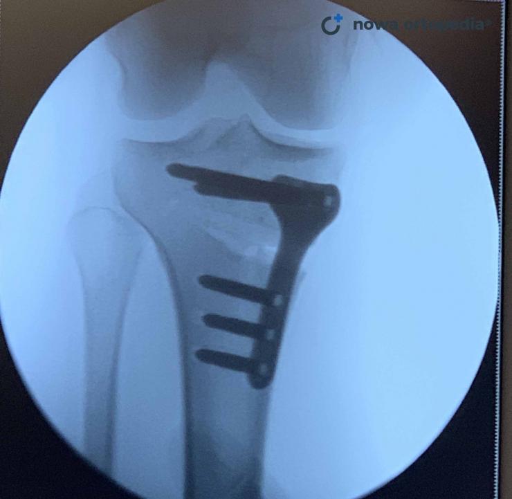
Result after 2 weeks - altered right limb axis 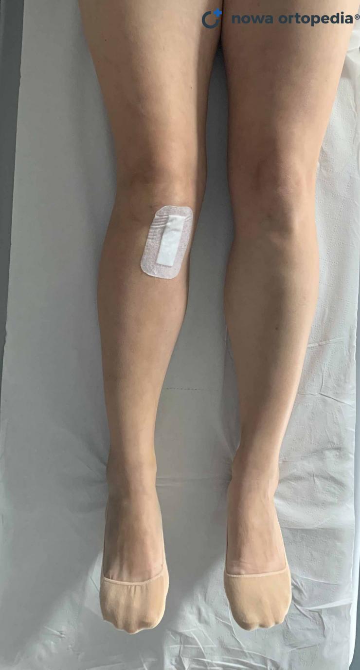
Closing popliteal osteotomy
In the past, the correction of the knee axis was performed mainly with the technique of lateral closing osteotomy. The surgeon makes an incision in the skin laterally and above the tibial tuberosity. Then, he crosses the tibia in two places at the right angle to cut a bone wedge with a predetermined base. The osteotomy gap is closed and stabilized with an anastomosis (e.g. with a plate). If necessary, it is possible to change the tension of the sagittal collateral ligament and the muscles attached to the head of the arrow on the side of the knee and to correct the tibial plateau forward inclination.
The tibia fixation obtained as a result of a closing osteotomy is very stable and allows for a rapid fusion of the tibia. The disadvantage of this technique is a greater risk of damage to the peroneal nerve and the difficulty of obtaining an accurate correction resulting from the need to perform two precise bone cuts and the occurring shortening of the limb. It is also harder to change the amount of tibia correction during surgery on an ongoing basis.
Open wedge
It is performed most often - from a medial approach. The surgeon makes an oblique incision of the tibia approximately 2.5 cm below the femoral tibial joint. The cut leaves about 10% of the width of the bone on the lateral side for stability. The fragments open, breaking the side of the bone. The height of the osteotomy gap, and thus the size of the correction, can be easily adjusted during the procedure, which is the advantage of the opening osteotomy over the closing one. The gap in the osteotomy can be filled with bone grafts taken from the patient, artificial grafts, or left unfilled. After its partial cutting, the bone is stabilized by a plate dedicated to this type of treatment - titanium or carbon fiber.
Open-wedge osteotomy of the varus knee has the following advantages:
only one incision of the tibia is required instead of two,
the possibility of more precise correction of the knee axis in both planes,
no bone loss in the proximal end of the tibia,
less interference in the structure of the tibiofibular joint,
lower risk of damage to the peroneal nerve,
easier parallel procedures on the knee, e.g. reconstruction of the anterior cruciate ligament or treatment of meniscus injuries,
the possibility of lowering or increasing the height of the patella by changing the cut line of the tibia - below or above the tibial tuberosity.
The disadvantages of opening osteotomy include:
higher risk of bone non-union,
greater risk of bone infection and septic loosening of the implant,
the need for a longer period of unloading the limb after the operation (but with a stable plate, we allow walking immediately).
Knee osteotomy - complications
Complications after corrective varus knee osteotomy are rare and include:
non-union or delayed union of the tibia (pseudo-joint),
too much or too little correction,
loss of correction despite correctly performed surgery,
thrombosis,
conflict between the bonding material and soft tissues.
Knee osteotomy - rehabilitation
The patient should be aware of the fact that properly conducted rehabilitation is as important as properly performed surgery. After the surgery, you should strictly follow the instructions of your doctor and physiotherapist. After the operation, passive movements of the knee on the CPM rail are introduced as soon as possible. During breaks between exercises, the knee joint can be cooled down by a special Game Ready device, thanks to which the pain sensations after the surgery are significantly reduced. The patient also performs isometric exercises of the quadriceps muscle and gradually introduces limited active knee movements. For the first few weeks after the operation, the patient moves on crutches with partial relief of the operated leg. The time when the limb can be fully loaded depends on the type of corrective osteotomy (open or closed), the type of anastomosis used, the quality of the bone tissue and possibly other parallel procedures performed in the knee. As a rule, the patient should use the crutches for 6-8 weeks after the procedure or until the fusion of the osteotomy fissure is detected on the X-ray.
In the process of physiotherapy after an osteotomy of a varus knee, it should be taken into account that the lower limb, and in fact the whole body, must undergo adaptation to new biomechanical conditions. The goals of the rehabilitation process will include:
restoration of a painless full range of motion in the knee joint,
postoperative scar mobilization,
mobilization of the kneecap and correct activation of the medial head of the quadriceps muscle of the thigh (VMO, vastus medialis obliquus) to improve the function of the patellar femoral joint,
learning the correct gait pattern with full loading of the operated leg after changing its angle and length,
functional strengthening of the muscles stabilizing the knee joint during everyday activities and sports activities.
Important information
| Duration of the procedure (depending on the method) | 40-120 minutes |
| Tests required for surgery | basic - preparation for surgery tab |
| Anesthesia | subarachnoid |
| Hospital stay | minimum 4-6 hours after surgery |
| Period of severe dysfunction (reduced amount of walking) | 2 - 3 weeks - about elbow crutches |
| A period of limited dysfunction | up to 6 weeks |
| Removal of stitches - first visit | 12-16 days |
| Change of dressings | every 3-4 days |
| Contraindications to the procedure | smoking, obesity, blood clotting disorders |
Frequently asked questions about the popliteal osteotomy procedure:
A popliteal osteotomy is based on the restoration of the correct mechanical axis of the knee joint thanks to the surgical correction of the proximal epiphysis of the tibia. This treatment causes a more even distribution of loads within the articular cartilage, which reduces the pain in the knee. A tibial osteotomy is an effective treatment for knee pain in young people with a disorder of the limb axis and in the early stages of osteoarthritis. In selected cases, tibial osteotomy is an alternative to the implantation of a knee joint endoprosthesis.
The most common indication for osteotomy surgery is pain in the knee joint of degenerative origin accompanied by varus of the tibia. Varus deformity of the tibia can be initially diagnosed when the knees are separated by a minimum of 4 cm with the joint ankle joints. A popliteal osteotomy makes it possible to correct the deformed axis of the limb and thus relieve the painful structures in the medial compartment of the knee.
The required rehabilitation time after a popliteal osteotomy is approximately 3 months. This time may also be shorter or longer depending on the individual anatomical conditions of the knee, procedures performed in parallel during the operation (e.g. reconstruction of the cruciate ligament) and the expected level of patient activity after the procedure.


