
Hand ultrasound is widely used in the diagnosis of chronic wrist pain, post-traumatic conditions, edema, inflammation and sensory disorders in the hand and fingers. The ultrasound examination most often assesses the joints of the hand, the structures lying in the carpal tunnel and the tendons of the fingers. Due to the possibility of dynamic tissue assessment, ultrasound is the most frequently chosen diagnostic method in most pathologies of the hand area. In some cases, ultrasound complements the results of functional tests, e.g. nerve conduction tests of the median nerve in carpal tunnel syndrome. Hand ultrasound, by locating pathological changes, facilitates the planning of surgical treatment. Moreover, ultrasound is an invaluable tool for monitoring the healing process and tissue reconstruction after interventional procedures, as well as for controlling the effectiveness of treatment of some pathologies, e.g. the condition after ganglion aspiration.
Make an appointment now - to the doctor who performs ultrasound of the hands in our hospital
[title]
[image-intro]
[readmore text="Read more"]{/article}
[title]
[image-intro]
[readmore text="Read more"]{/article}
Wrist ultrasound
Ultrasound of the dorsal side of the wrist
The wrist ultrasound examination on the dorsal side includes the assessment of the structures lying under the so-called cord of rectifiers. During the examination, the doctor assesses the muscle tendons located in the various compartments (I-VI) of the reticulum:
1st compartment: tendons of the abductor muscles of the long thumb and the extensor muscles of the short toe,
2nd compartment: tendons of the muscles of the radial extensor of the long wrist and the radial extensor of the short wrist,
3rd compartment: long extensor muscle tendon of the thumb,
4th compartment: tendons of the extensor muscles of the fingers and the extensor muscles of the pointer,
V compartment: extensor tendon of the little finger,
VI compartment: wrist extensor tendon.
Then the following are assessed:
wrist bones with ligaments,
bone outlines of the base of the distal bones of the forearm,
ligaments of the radiocarpal joint,
Triangular Fibrocartilage Complex (TFCC) - it is a cartilage-ligament tissue located between the wrist bones and the ulna.
Ultrasound of the palm side of the wrist
The wrist ultrasound examination on the palmar side includes the assessment of:
structures located in the carpal tunnel:
median nerve,
finger flexor tendons,
structures located in the Guyon Canal:
ulnar nerve,
the ulnar artery.
What pathologies can be diagnosed during ultrasound of the hand?
De Quervain's disease
A symptom of de Quervain's disease is pain at the base of the thumb and the side of the wrist. The essence of the disease is inflammation of the tendon sheaths of the muscles that straighten and abduct the thumb. These tendons run in the first compartment of the extensor cord. Reducing the space under the strap and overloading the thumb muscles can lead to mechanical tissue irritation.
Ultrasound examination is the basic method of imaging de Quervain's disease. During the ultrasound examination, the doctor assesses the structure of the tendons, the surrounding sheaths and the extensor cord. A dynamic ultrasound examination allows for the observation of abnormalities in the sliding of the tendons in the sheath during thumb movements. The physician often finds thickening of the tendons above and below the strap and the pulling of the fibrotic cord through the tendons of the muscles.
Read more about de Quervain's disease.
Crossroads syndrome
Symptoms of cross-border syndrome are pain and swelling in the distal part of the forearm on the radio-dorsal side. In the course of the cross syndrome, tendon sheath inflammation occurs due to the mutual conflict of the intersecting tendons of the muscles belonging to the first and second extensor retinitis compartments. Crossover syndrome often occurs in people who perform repeated movements of flexing and extending their wrists.
Ultrasound shows exudation and thickening of the tendon sheaths, and within the tendons, fibrosis and areas of microdamage are found.
Wartenberg's disease
In the course of Wartenberg's disease, pain and tingling of the radial-dorsal side of the wrist and the distal part of the forearm are observed. The cause of the ailments is irritation of the radial nerve branch due to inflammation of the adjacent cephalic vein, pressure by the fascia of the forearm or scarring lesions.
Ultrasound allows for a precise assessment of pathological conditions potentially causing a conflict with the radial nerve branch. Ultrasound also shows post-traumatic nerve disruption. The results of the ultrasound examination are crucial for the selection of the treatment method.
Ganglions
Ganglions (gelatinous cysts) are soft, fluid-filled bumps located on the dorsal side of the wrist. Ganglions are probably formed as a result of damage to the wrist capsulo-ligament apparatus, which causes the fluid to escape outside the joint cavity.
Wrist ultrasound allows you to locate ganglions and differentiate them from other benign tumors in the hand, such as a tendon sheath tumor (giant cell tumor) or lipoma.
Read more about ganglion treatments.
Carpal tunnel syndrome
Carpal tunnel syndrome manifests itself as numbness or pain in the palmar side of the wrist radiating towards the 1st-3rd fingers and the middle of 4th finger. Ailments result from compression of the median nerve at the level of its passage through the carpal tunnel.
Carpal tunnel ultrasound enables the assessment of the median nerve and the canal structures that may be the reason for the reduction of the nerve space and its compression. Possible causes of pressure include: flexor tendon sheath inflammation, traumatic scarring, degenerative changes or the presence of ganglions. In pathological conditions, the doctor finds the rounded shape of the nerve in its cross-section, decreased echogenicity, obliteration of the fibril structure of the nerve and local increased blood circulation causing the nerve swelling.
Read more about carpal tunnel syndrome.
Guyon's Channel Syndrome
The ulnar nerve and the ulnar artery run in the Guyon canal formed by the pinhole and the hook bone. Compression of the ulnar nerve triggers sensory disturbances or pain radiating to the 4th and 5th fingers. The cause of pinching the nerve may be repeated mechanical compression while resting your wrist frequently on the crutch holder or the handlebars of a bicycle. Most often, however, the nerve is pinched by post-traumatic swelling of the soft tissues, a bone fragment, the presence of a tumor or an aneurysm in the ulnar artery. Hand ultrasound examination allows for precise visualization of the cause of nerve compression.
Sonosurgery
Sonosurgery is a percutaneous intervention performed under ultrasound guidance. Examples of sonosurgery within the hand are:
Wrist and hand joint puncture - collection of synovial fluid,
Administering the drug to the wrist and hand joints,
Administration of the drug into the inflamed tendon sheath,
Aspiration of ganglion content.
A hand ultrasound may also include an ultrasound of a finger.
Source:
- Dębek A, Czyrny Z, Nowicki P. Zmiany patologiczne ręki w badaniu ultrasonograficznym. Journal of Ultrasonography 2014; 14: 74-88.
- Bianchi S, Zamorani MP. Procedury interwencyjne [w:] Derchi L, Rizatto G, Valle M, Zamorani M. Ultrasonografia układu mięśniowo-szkieletowego, Medipage, Warszawa 2009, s. 889-918.
Frequently asked questions about ultrasound of the wrist, hand:
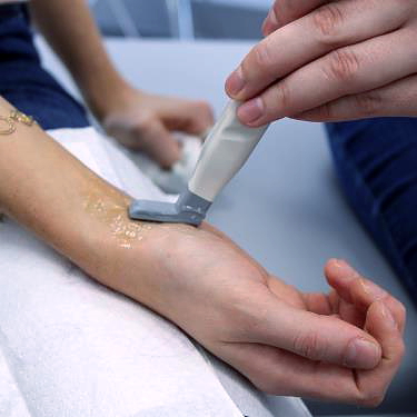
During the ultrasound examination, the doctor places a special probe dedicated to the examination of small joints and smaller anatomical structures of the body to the dorsal or palmar surface of the wrist, hand or fingers. The scope of the examination depends on the indications resulting from the clinical picture. Ultrasound examination of the hand allows for the identification of damage to muscle tendons, inflammation of the tendon sheaths and joints, as well as degenerative and productive changes. Thanks to the ultrasound examination, it is possible to assess the course of the median and radial nerves and the possible cause of compression of these nerves.
The ultrasound examination may be performed by a radiologist, orthopedist or rheumatologist with a certificate of training in ultrasound of the wrist and hand structures.
Hand ultrasound does not require any special preparation. It is worth bringing the results of other tests, e.g. X-rays of the hand, laboratory results for rheumatoid diseases or the results of nerve conduction test in case of suspected compression neuropathy.
The duration of hand ultrasound depends on the range of structures assessed. In the case of rheumatological changes, the wrist and each finger are assessed separately, which may extend the duration of one hand examination to 50 minutes. When clinical examination indicates the need for an ultrasound examination of a single structure, e.g. the median nerve in the carpal tunnel, the ultrasound usually takes about 10-15 minutes.


