
Anterior cruciate ligament (ACL) injury is one of the most common injuries in the knee joint. Surgical treatment of the torn ligament is performed using the knee arthroscopy technique - a minimally invasive procedure involving the insertion of the camera and surgical instruments into the joint through small incisions. Most often, in the case of a total old rupture, the operator replaces the damaged ligament with a biological graft (or less frequently with a synthetic prosthesis). The anterior cruciate ligament reconstruction procedure allows to restore the knee stability required not only for safe sports or physical work, but also reduces the risk of faster development of degeneration and subsequent injuries of the joint with meniscus damage.
Make an appointment now - with a doctor specializing in ACL reconstruction at our hospital
[title]
[image-intro]
[readmore text="Read more"]{/article}
[title]
[image-intro]
[readmore text="Read more"]{/article}
ACL function of the anterior cruciate ligament
ACL is an intra-articular ligament. It begins with a narrow longitudinal attachment on the medial surface of the lateral femoral condyle, runs obliquely downwards medially and ends with a lunar-shaped attachment on the anterior intercondylar field of the tibia.
The main role of the anterior cruciate ligament is to inhibit the anterior displacement of the tibia in relation to the femur and to ensure rotational stability of the knee joint. In addition, the cruciate ligament is extremely rich in deep feeling receptors, the so-called proprioreceptors. Information about the tension of the ligament is transmitted to the central nervous system, on the basis of which the response is programmed on an ongoing basis in the form of activation of appropriate muscles that actively stabilize the knee joint. The ACL therefore plays an important role in creating a knee-safe movement pattern.
ACL anterior cruciate ligament injury
The anterior cruciate ligament in the healthy knee can withstand a force of about 2500N, although the ACL strength may be lower in elderly people with severe knee degeneration. While walking, the ACL ligament exerts a force of about 400N, and during sports activities related to running, making sudden turns and jumps, it can increase to about 1700N.
The anterior cruciate ligament ruptures when the force acting on the ligament exceeds its mechanical strength.
Usually, along with ACL rupture, other structures of the knee are damaged, including most often the lateral or medial meniscus, medial colateral ligament (MCL), and antero lateral ligament (ALL).
The most common mechanism of ACL damage is the combination of knee rotation with valgus. This situation may occur:
during rotation, when the thigh follows the body and the tibia remains fixed by the foot on the ground,
as a result of a direct injury - a hit to the knee of a loaded limb (e.g. in contact sports),
during landing (jumping off) on the poorly stabilized muscular lower limb.
ACL fault diagnosis
The severity of symptoms depends on the degree of ligament damage:
I - straining of the ligament, a small number of fibers have been damaged; knee stability is maintained,
II - damage to a larger number of fibers, post-traumatic instability of the knee joint not greater than 5 mm (compared to the opposite healthy limb),
III - complete rupture of the ligament, detachment of ligament attachments from the bone or excessive stretching of the ligament resulting in instability of the knee joint greater than 5mm.
At the time of complete rupture of ACL (Grade III), severe pain, sometimes audible crackling, may occur. Within hours of the injury, pain and swelling gradually increase. Additionally, there is a feeling of insecurity and the knee running away when trying to load the leg with body weight.
As part of the physical examination, the orthopedic surgeon performs clinical tests, such as the anterior drawer test, Lachman test, or Pivot Shift, on the basis of which he can initially determine an anterior cruciate rupture.
A very common symptom in ACL ruptures (it is estimated that in over 80% of ruptures) is the presence of a hematoma in the knee.
Examination of the knee after injury is almost always biased - pain and reflex muscle tension make it impossible to perform basic tests assessing ligament efficiency. Therefore, the examination of choice is high-field magnetic resonance imaging of the knee (MRI)
This does not change the fact that the most frequently performed examination after a knee injury in the HED (Hospital Emergency Department) is X-ray, in which the so-called Segond fracture is sometimes observed - a detachment of a small bone fragment on the lateral side of the tibia (corresponding to the damage to the ALL mentioned). This symptom is considered a radiological synonym for an ACL rupture.
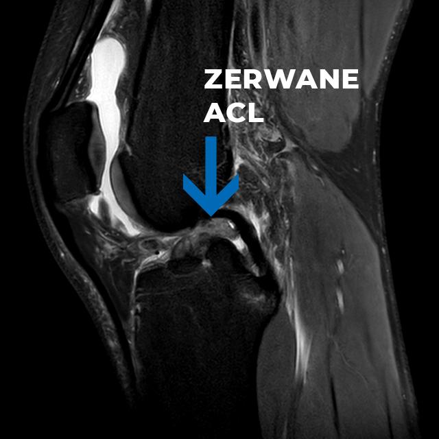
MRI image 5 days after breaking the ACL
Long-term effects of ACL breaks
The lack of efficient ACL may lead to disturbances in the mechanics of the knee joint, which are felt by the patient mainly during sports and physical work. The reported symptoms are the feeling of the knee escaping and chronic pain resulting from secondary overloads of the remaining knee structures. Under conditions of joint instability, the risk of meniscus damage and recurrent knee swelling increases. Failure to treat traumatic instability accelerates the development of osteoarthritis of the knee (gonarthrosis).
Indications for arthroscopic ACL reconstruction
The purpose of ACL reconstruction is to improve the comfort of life by restoring the mechanical stability of the knee joint.
ACL reconstruction is most often performed in people:
under 55 years of age,
who underwent a conservative treatment process that did not end with restoration of knee stability,
who plan to increase the level of activity, do recreational sports,
professional athletes,
with ACL injury in both knee joints.
Preparation for surgery - rehabilitation
Appropriate rehabilitation preparation allows to improve the functional state of the limb even before the planned surgery, which significantly affects the result of the ACL reconstruction procedure, and also facilitates the implementation of targeted physiotherapy methods after the procedure. As a result, recovery from surgery will be faster.
Rehabilitation before the planned ACL reconstruction surgery will be aimed at:
elimination of intra-articular exudate, swelling and knee pain,
prevention of contractures and restoration of a painless full range of motion in the joint,
obtaining optimal tension and restoring the strength and endurance of the muscles of the lower limb,
improvement of deep feeling, stimulation of equivalent reactions.
The course of reconstruction of the anterior cruciate ligament
The surgical procedure is performed under subarachnoid anesthesia blocking the feeling below the waist - the patient remains conscious during the operation and can observe the course of the procedure on the monitor screen. The surgeon introduces the arthroscope and working tools through small incisions in the knee, with which he assesses the damage to the ACL ligament and other joint structures (posterior cruciate ligament, meniscus, articular cartilage, synovium). It removes pathological changes that may be the cause of knee pain and repairs damaged structures, e.g. it supplies defects in articular cartilage and sutures the meniscus, which often breaks simultaneously during a knee injury leading to ACL rupture.
In most cases (except for the Internal Bracing method), the surgeon also removes the remains of the damaged anterior cruciate ligament. Then he prepares a transplant that can be taken from the patient (autologous transplant), a transplant from a deceased donor (allograft) or a synthetic ACL prosthesis.
The next step is to drill the bone tunnels in the femur and tibia where the graft will be placed. The new ligament is inserted into the joint and stabilized in the bone so that its course and tension are as close as possible to the anatomical ligament.
The method described above is the most standard, classic way of treating an unstable knee.
A modern approach to the reconstruction of the anterior cruciate ligament
In the last 10 years, due to a wider understanding of the functioning of the knee joint (in terms of biomechanical and biological points of view), as well as greater knowledge resulting from the conclusions drawn from the observation of patient outcomes, there has been a significant diversification of the procedures.
We currently use treatments aimed at preserving our own ligament as much as possible (mainly due to its unique ideal attachment area, vascularization, innervation and attachment potential) and treatments using additional internal splinting to prevent ligament elongation during its reconstruction.
The approach to when to reconstruct the anterior cruciate ligament has also changed.
Until now, early reconstructions have been avoided by attempting to heal a stable knee conservatively in a cast or orthosis.
Currently, thanks to the possibility of quick MRI diagnostics, we believe that the best approach is arthroscopy of the knee in the period not later than 6 weeks after the injury, in which we try to reinsert ACL on a titanium anchor or suture ACL with the simultaneous performance of the Internal Bracing technique.
It is especially possible in the case of ACL detachment from the femur, and much more difficult in the case of intra-segmental ligament injuries.
Sometimes, intraoperatively, we make a decision to perform a partial (hybrid) ACL reconstruction - i.e. to perform a small ACL graft from only 1 tendon and simultaneous attachment of the ACL stump remaining after rupture to the femur at the site of the graft implantation. This results in much better proprioception ("knee feeling") and vascularization of the reconstructed ligament.
Internal Bracing
Internal Bracing is a modern method supporting the repair of a broken ACL. It can be used as an additional reinforcement added to the classic ACL reconstruction - this is how we always perform ACL reconstructions in our hospital.
It can also be a technique supporting reinsertion, i.e. sewing a torn cruciate ligament to the femur.
Inetrnal bracing, or in free translation: "internal splinting" is a method where we use a strong non-absorbable 2 mm thin polyester tape to secure the distance between the femur and tibia during the healing of the grafted or the growth of the sewn anterior cruciate ligament.
The tape is not removed.
Internal Bracing used for ACL reinsertions should be performed within 6 to 8 weeks after the injury. Later, the ligament will no longer be able to attach itself to the femur. In the meantime, it shrinks and deforms when the knee is in use.
The advantages of the Internal Bracing method include:
preservation of your own ligament and no need to collect a graft (during reinsertion)
lower degree of impairment of deep sensation,
preservation of the natural area of attachment of the ligament to the tibia, positively influencing the biomechanics of the knee,
less traumatization of tissues - no need to drill 7-9 mm tunnels in the femur and tibia as in classic reconstruction,
shorter recovery time - after 3-4 months after surgery, the endurance of the healed ACL is equal to the baseline value from before the injury.
performing this type of procedure does not close the way to complete ACL reconstruction in the future in any standard way (completely different than when the patient has already had a full ligament reconstruction performed earlier)
One-bundle and two-bundle ACL reconstruction
Classic complete reconstruction of the anterior cruciate ligament involves carrying out the graft through the knee joint in the form of a single bundle. For several years, there has been a discussion about the two-bundle structure of the natural ACL ligament and the importance of this fact for obtaining the most similar biomechanics of the knee during reconstructive surgery. These discussions resulted in the concept of ACL two-bundle reconstruction, in which two separate graft bundles are attached at different locations on the tibia and femur. To implant new ACLs in this way, however, it is necessary to drill an additional two bone tunnels. The superiority of the two-bundle procedure over the one-bundle procedure requires confirmation by a larger number of independent studies. The most important for the success of the procedure is the operator's experience, knowledge of biomechanics and proficiency in adapting surgical techniques to the patient's specific knee joint.
Types of transplants
The mechanical strength of the graft decreases during the healing process (6-12 weeks after the procedure), therefore the initial force that the graft can withstand must be higher than that of the natural ACL. ACL transplantation goes through the stages of:
ischemic necrosis,
revascularization,
redevelopment.
The ligamentization process (transformation of the transplanted tissue into an efficient structure replacing ACL) is long - it lasts 8-12 months and depends on the type of the harvested graft, the method of its fixation, individual patient characteristics and many other factors.
Hamstrings of the hamstrings
The most frequently chosen type of transplant is the semi-tendon and slender tendon. They are taken from the lower-medial part of the knee joint. The obtained tendons are folded in four ways to obtain better mechanical strength and are implanted on bioabsorbable screws or screws, plates or bolts made of titanium. The hamstring graft is a good choice for people for whom the removal of the patellar ligament is inadvisable, e.g. due to the lateral support of the patella or the presence of significant degenerative changes in the patellofemoral joint.
In our hospital, we most often use the method of fixation on the tibia with the use of endobutton (a type of button). This allows for the quadruple assembly of only 1 tendon and therefore the performance of a full-fledged ACL reconstruction without the need for 2 hamstrings.
Patella ligament
The graft is the central band of the ligament together with the bone blocks. This allows the attachments of the very strong patellar ligament to be firmly attached to the bone. The disadvantage of this method is the risk of complications in the form of knee extension apparatus disorders: excessive tension of the rectus muscle of the thigh, scarring limiting knee flexion, the possibility of kneecap fracture and the development of chronic pain in the patellofemoral joint. Currently, based on many years of observation, it is believed that it is not the ligament strength but the correct location of the graft that is most important for its long-term proper functioning.
Quadriceps hamstring
During the procedure, the central part of the quadriceps tendon is collected together with the patellar bone block. This method is often used during revision reconstructions in people who have already received an autologous transplant.
Transplant from a deceased donor (allograft)
ACL reconstruction can also be performed using a transplant obtained from a deceased donor (from a tissue bank) - most often it is the patellar ligament or the Achilles tendon. This avoids the inconvenience of collecting the patient's own tissues. The disadvantage of this method is the price and the negligible but existing risk of infection and healing in the ligament elongation.
LARS - ACL synthetic prosthesis
LARS prostheses completely replace the knee ligament, so there is no need to get your own graft. An artificial ACL implant is implanted in bone tunnels and stabilized with an appropriate anastomosis. Returning to normal activity and sport is much faster (the first trainings are possible even in the second month after the treatment). The downside of this method is a greater risk of avulsion fractures of attachment of the prosthesis to the bone, the risk of infection and bone lysis around the graft. Calibration for the LARS implant procedure must be very careful in consultation with the patient and should take into account the individual biomechanical conditions of the knee and the type of sport practiced. We avoid the reconstruction of a completely artificial ligament whenever possible.
Rehabilitation after ACL reconstruction
Rehabilitation begins on the first day after the operation and consists in gradual upright standing and exercises of active limbs outside the operated one. The physiotherapist supports the patient when moving on crutches and teaches how to put on an orthosis. Supporting the gait with crutches is helpful for up to 3 weeks after the procedure. The orthosis should be put on for activities where there is a risk of uncontrolled knee movement, but most often it is put off in the 4th week after the procedure. Knee exercises that improve their active stabilization are of key importance for the protection of the graft.
The main goals and tasks that rehabilitation after ACL reconstruction should fulfill are:
Minimizing pain and swelling after surgery
Discomfort and slight swelling occur after each knee surgery. From the point of view of rehabilitation, this condition is unfavorable because it leads to a reflex increase in muscle tension and makes it difficult to introduce exercises to restore mobility in the joint. The Dworska Hospital uses a special Game-Ready device that allows simultaneous cooling and optimal compression of the operated knee. The effect of these activities is a better well-being of the patient and the possibility of faster implementation of exercises after the procedure.
Restoration of full range of motion in the knee
After the operation, we strive to achieve full extension of the knee as soon as possible. The extent of flexion depends on the type of graft and the method of its fixation, but in most cases, aim to achieve 90 degrees of flexion as early as 7-10 days after ACL reconstruction. By the end of week 8, the flexion range should be 135 degrees without attempting to exceed it, as this could stretch the graft. The complete safe range of knee mobility is achieved 12 weeks after the procedure.
Postoperative scar mobilization
The scars at the places where the arthroscope and surgical instruments are introduced are small (up to 1 cm) and heal quite quickly. A bigger problem can be caused by scarring in the areas where the graft was taken - the lower-medial surface of the knee (the tendon of the semitendinus or slender muscle) and the front of the knee (the patella ligament). After removing the stitches (10-14 days after the operation), manual preparation of the scar should be started. The therapy can be supported with kinesiotaping and physical therapy treatments. Failure to act in this direction may result in the formation of a clearly visible, inelastic scar that painfully restricts the range of motion.
Rehabilitation taking into account the place of harvesting the transplant
Pain at the site of the graft collection is a frequent problem following the reconstruction of the anterior cruciate ligament with the use of own tissues. Rehabilitation will aim to improve the function of the patellofemoral joint, the quadriceps muscle of the thigh or the hamstring muscles.
Improving deep feeling
Damage to the natural ACL ligament is associated with the loss of the deep feeling receptors located in it. The knee after surgery with an implanted ACL implant will have a limited ability for automatic muscular stabilization. Proprioception training that stimulates the mechanoreceptors of the remaining knee structures is crucial for the prevention of graft failure in the future.
Proper activation and muscle strengthening
An important task of rehabilitation is the activation of the medial head of the quadriceps muscle of the thigh (VMO, vastus medialis obliquus), which is crucial for the functional stability of the knee. This muscle weakens the earliest after surgery and often ceases to be properly activated. The therapy in the first weeks after the procedure can be supported by electrostimulation of the VMO muscle - especially in those patients who have difficulties with activating this muscle on their own.
Another task of physiotherapy after ACL reconstruction is to strengthen the gluteal muscles and the muscles of the anterior and posterior thigh groups. According to some concepts, it is recommended to obtain an appropriate relationship between the strength of the quadriceps muscle (extending the knee) and the strength of the ischio-shin muscles (bending the knee).
During rehabilitation, a limiting factor should be taken into account in the form of a decrease in the strength of the natural graft between the 6th and 12th weeks after the procedure. Rehabilitation exercises up to the 12th week after the procedure should be performed in a closed kinematic chain - with a stabilized foot resting, for example, on a wall or floor. Performing this type of exercise facilitates the simultaneous activation (co-contraction) of the opposing muscle groups, which allows them to strengthen while minimizing the anterior shear forces that can stretch the maturing graft.
Functional training
The most important goal of rehabilitation after ACL reconstruction is to recreate the knee stability mechanism during movement - so that the knee muscles act as a dynamic stabilizer during functional patterns, e.g. walking, going up and down stairs, getting on and off the bus or jogging. The aim of physiotherapy will be to obtain the correct gait pattern and all activities of daily living that the patient undertakes.
Back to sport
We introduce exercises specific to a given sport discipline when increasing the intensity and difficulty of the exercises does not cause pain or swelling in the knee, and the results of functional tests of the operated limb are at least 80-85% of the healthy limb result. Return to light sports activities is possible 6-7 months after the operation. Heavy-load training and participation in contact sports or sports that require rotational stability (football, basketball, skis) can be started about a year after the ACL reconstruction.
Important information
|
Duration of the procedure (depending on the method) |
50 - 120 minutes |
| Tests required for surgery | basic - preparation for surgery tab |
| Anesthesia | subarachnoid or general |
| Hospital stay | 6 - 12 hours |
| A period of significant dysfunction | 2 - 3 weeks |
| A period of limited dysfunction | 4 - 12 weeks |
| Removal of stitches - first visit | 12 - 16 days |
| Change of dressings | every 3 - 4 days |
| Contraindications to the procedure | infection, 3rd and 4th degree degeneration of the knee |
Frequently asked questions about ACL ACL reconstruction surgery:
The ACL reconstruction procedure is performed when severe knee instability (3rd degree) is found, which makes it difficult for active people to function normally. The aim of the treatment is to improve the quality of life and to prevent the rapid development of knee osteoarthritis. Surgical ACL reconstruction is especially recommended for athletes and blue-collar workers. Less degrees of ACL damage and complete rupture of the ACL ligament in people leading a sedentary lifestyle can be successfully treated conservatively through specialized rehabilitation.
The length of treatment depends on many factors, including: rehabilitation of the patient before the procedure, the type of transplant used (own, donor or artificial implant), the presence of additional damage to the knee and the patient's involvement in the physiotherapy process after the procedure. In the case of the most frequently performed ACL reconstructions with the use of own muscle tendons or the patellar ligament, the time to return to normal activity is about 3 months. You can start practicing sports in the period from 6 to 12 months after the procedure.
The first steps after the procedure can be taken as early as 1-2 days after the surgery. During the first 3-4 weeks after the procedure, walking is possible with the support of the elbow crutches and with the knee brace. Free walking with full load on the limb without additional support is possible when the physiotherapist determines that the knee is fully prepared and trained for it, and moreover, walking does not cause recurrence of swelling or pain in the joint.
Rehabilitation begins on the first day after surgery and should be systematically continued under the supervision of a qualified physiotherapist for at least 3 months. Properly implemented treatment will allow you to restore the full range of motion in the knee in the optimal time, regain the required muscle strength and stability of the joint, and as a result will allow you to safely return to normal activity and sport.


