What is the purpose of ultrasound examination in rheumatology?
Ultrasound is an auxiliary tool in order to make a correct diagnosis of the disease. It allows you to immediately visualize the changes in the affected joints during the visit and to determine whether there is active inflammation, which is of great importance in choosing the right treatment. It enables the detection of early inflammatory changes that precede joint destruction. It also allows you to monitor the therapy and confirm the remission of the disease, i.e. the transition of the disease into an inactive process. Thus, it facilitates the decision to change treatment or reduce the doses of medications taken.
The ultrasound also allows for precise administration of drugs into the joint cavity, tendon sheaths or bursae and collection of synovial fluid for examination.
Make an appointment now - to the doctor who performs ultrasound of joints in our hospital
[title]
[image-intro]
[readmore text="Read more"]{/article}
Who performs rheumatological ultrasound?
The most common indication for an ultrasound examination is pain and swelling in the joint.
Rheumatological ultrasound should be performed when an inflammatory disease is suspected and an appropriate diagnosis cannot be made on the basis of an interview, physical examination and additional examinations. One swollen joint is enough, accompanied by pain or morning stiffness that was not caused by trauma or overstrain. Especially when a greater number of joints are involved and it is accompanied by general symptoms, such as low-grade fever or fever, weakness, weight loss. Also, in people with already diagnosed rheumatic diseases, an ultrasound is performed to assess the effectiveness of the treatment.
Compared to X-ray examination, it is safe, it does not expose it to radiation, it allows to visualize the active inflammatory process that takes place in structures that cannot be distinguished on a classic X-ray image. The ultrasound allows you to visualize the changes earlier than the X-ray examination.
What pathological changes can be visualized in ultrasound in rheumatic diseases?
Ultrasound is used to assess the structures of the musculoskeletal system that may be inflamed in the course of systemic diseases - this applies to virtually all peripheral joints, i.e. the shoulder, elbow, knee, ankle, hip, wrist, hand, toe and foot joints. .
The ultrasound image is not specific to a specific disease, but it allows to visualize a number of inflammatory and post-inflammatory changes.
During the examination, individual articular structures (bone articular surface, articular cartilage, articular cavity, synovium, bursae) and periarticular structures (ligaments, tendons, muscles, nerves) are assessed. Each of the replaced elements is assessed in terms of the presence and advancement of pathological changes.
The changes that may indicate rheumatic diseases include:
- the presence of fluid in the articular cavity and its recesses
- changes in the synovial membrane - its hypertrophy, hyperemia or fibrosis
- the presence of erosions
- tendon pathologies - enthesopathies
- inflammation of tendons and tendon sheaths
- changes in articular cartilage
- bursitis
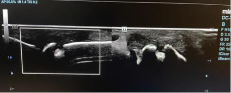
Figure 1 erosion in the metacarpophalangeal joint
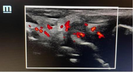
Figure 2 Exudate and synovitis in the metacarpophalangeal joint
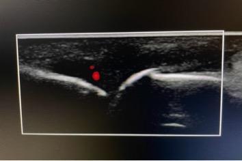
Figure 3 Exudate in the metacarpophalangeal joint II
The Power Doppler vascular function is extremely useful in ultrasound in rheumatology, showing increased vascularization of the examined structure, and thus the presence and severity of the inflammatory process.
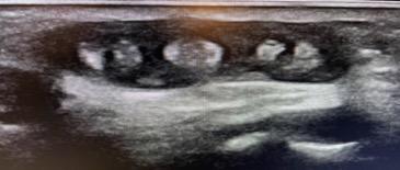
Figure 4 The effusion around the extensor tendons of the fingers
In what rheumatic diseases is ultrasound useful
Early arthritis
In the diagnosis of rheumatological diseases, it is of key importance to quickly diagnose the disease and implement appropriate treatment in order to halt the progression of the disease early and reduce the risk of irreversible changes. Ideally, the therapy should start within 3 months ("window of opportunity").
With minor clinical symptoms, at the beginning of the inflammatory disease, USG allows to distinguish intra-articular exudate from periarticular soft tissue edema.
Ultrasound examination of joints allows for quick detection of inflammatory changes in painful joints, as well as in joints and periarticular structures that do not yet show any symptoms or changes in physical examination, and in which the inflammatory process is already underway in the course of systemic disease. It allows you to visualize a small amount of exudate that cannot be determined by a clinical examination. Finding characteristic changes in ultrasound plays an important role in the differential diagnosis, it can predict the progression to the full-blown form of the disease and enables faster diagnosis and implementation of appropriate therapy.
Rheumatoid arthritis
The first changes in the ultrasound in the course of RA are synovial hypertrophy with concomitant exudate in the joint cavity, tendon sheaths or bursae. When the inflammatory process is active it may show increased synovial vascularization in the power Doppler. Ultrasound examination allows to distinguish whether the pain is associated with active inflammation or irreversible destructive changes, which is of key importance for the choice of treatment. The imaging of bone erosions is also of particular importance in confirming the diagnosis of the disease. The presence of such changes in typical places can confirm the presence of the disease and is one of the indicators of its aggressive course. Ultrasound allows you to visualize erosions at a much earlier stage than X-rays, which allows you to quickly apply appropriate treatment.
Spondyloarthropathies - psoriatic arthritis, ankylosing arthritis, inflammatory bowel disease, reactive arthritis
Spondyloarthropathies are a group of diseases in which the axial joints (sacroiliac joints and spine joints), peripheral joints, tendon attachments and changes in many other systems and organs are affected. The most common ultrasound examination is used to detect tendon enthesitis, the so-called entesitis. It mainly concerns the Achilles tendon attachment, plantar fascia and patellar ligament, quadriceps attachment, lateral epicondyle attachments and finger flexor tendon attachments. The ultrasound examination shows a thickening of the tendon, a decrease in its echogenicity and an increase in Power Doppler flow, it is also possible to visualize permanent changes in the form of calcifications. Early diagnosis and initiation of anti-inflammatory treatment and physiotherapy inhibit the formation of irreversible changes. In the course of spondyloarthritis, one or more peripheral joints are most often affected - primarily the knee, ankle, elbow, and in the case of psoriatic arthritis - the joints of the hands, feet and wrist. This can be confirmed by ultrasound examination with synovitis. In addition, the inflammatory process may affect tendons, tendon sheaths (primarily the flexors and extensors of the hands) and bursae (mainly the Achilles tendon bursa). Psoriatic arthritis often causes swelling and inflammation of the whole finger ("sausage finger") - dactylitis, which can also be visualized on ultrasound.
Crystallopathies - gout, chondrocalcinosis, hydroxyapatite crystallopathy
It is a group of diseases caused by crystallization and deposition in joints and periarticular structures of sodium urate, calcium pyrophosphate or hydroxyapatite crystals.
The examination confirming the disease is the detection of crystals in the synovial fluid, however, ultrasound examination may direct further diagnosis and help in the quick diagnosis of the disease.
The characteristic changes in an acute attack of gout include thickening and hyperemia of the synovium of the joint cavities, sheaths and bursae, and joint effusion. In chronic arthritis, changes in the articular cartilage (a characteristic symptom of a double contour), tophus, erosions, and deposits in the tendon attachments and ligaments can be visualized.
The most typical for chondrocalcinosis are calcifications in the hyaline cartilage (mainly in the knee joint and the head of the humerus).
In the rarely diagnosed hydroxyapatite arthropathy, caused by the accumulation of calcium phosphate crystals in the tissues, it is possible to visualize deposits in the tendon attachments and periarticular structures, most often large joints - the shoulder and the knee.
Rheumatic polymialgia
The changes that can be visualized in the course of rheumatic polymyalgia are included in the criteria for the diagnosis of the disease. Belong to them:
- bursitis
- inflammation of the tendon sheath of the long head of the biceps (tenosynovitis)
- inflammation of the shoulder joint
- inflammation of the hip joint
- trochanteric bursitis
Osteoarthritis
In the course of degenerative disease, the articular cartilage is damaged, which leads to secondary changes in the bones and in the synovium. The ultrasound examination shows the effusion in the joint cavity, a thinner layer of articular cartilage and osteophytes resulting from repair processes.


