CONTACT: contact@dworska.pl | +48 12 352 25 25

Mezoterapia stanowi niechirurgiczny zabieg medyczny z zakresu medycyny estetycznej oraz dermatologii, mający na celu dostarczenie skórze niezbędnych substancji odżywczych. Zabieg ma charakter profilaktyczny i leczniczy jednocześnie, gdyż pomaga w regenracji skóry na poziomie warstw głębokich.

photo 1509967419530 da38b4704bc6

Wskazania do wykonania zabiegu

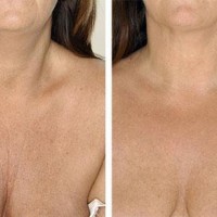
Zabieg zalecany jest w przypadku istnienia widocznych zmian skórnych oraz problemów o podłożu dermatologicznym np. blizn po trądzikowych. Mezoterapia pomaga odnowić właściwy poziom nawilżenia i napięcia skóry, dzięki czemu wspomaga walkę o idealnie gładką skórę. Zalecana jest jako zabieg redukujący zmarszczki w obrębie twarzy, szyi oraz dekoltu. Dzięki zabiegowi można zredukować widoczne blizny na całym ciele i cienie pod oczami. Mezoterapia niweluje rozstępy oraz cellulit, szczególnie w partiach ciała, jak pośladki, uda czy brzuch. Mezoterapia stosowana jest zarówno w przypadku defektów kosmetycznych, jak i przy konieczności odżywienia i nawilżenia głębokiego skóry w celu zredukowania oznak zmęczenia czy starzenia.

Zabieg można przeprowadzić niezależnie od wieku oraz kondycji skóry pacjenta. Śródskórne wprowadzenie substancji aktywnych zapewnia szybką regenerację skóry.

Przebieg procesu

Zabieg opiera się w początkowej fazie na zdefiniowaniu defektów kosmetycznych przez lekarza specjalistę oraz ich stadium zaawansowania. Zabieg można przeprowadzić na każdym etapie rozwoju defektów kosmetycznych. Lekarz po szczegółowej analizie kondycji skóry dobiera odpowiedni środek do indywidualnych potrzeb pacjenta, a następnie wstrzykuje go bezpośrednio w niewielkich dawkach w skórę w miejscach podlegających zabiegowi. Podanie substancji aktywnych podskórnie sprawia, że efekty rewitalizacyjne są szybsze i widoczne po krótszym czasie. Najpopularniejszymi preparatami, używanymi do zabiegu mezoterapii są środki m.in. Juvederm, Croma, Restylane, Teosyal, Fillmed.

Przeciwwskazania do wykonania zabiegu

Jak w przypadku każdego zabiegu z zakresu medycyny (także estetycznej) istnieją przeciwskazania do podjęcia zabiegu, a wśród nich w pierwszej kolejności wymienia się ciążę ze względu na brak informacji odnośnie negatywego wpływu terapii. Osoby, które z obniżoną krzepliwością krwi powinny zrezygnować z mezoterapii. Zabieg odradza się również chorym na zakażenia bakteryjne skóry tj. trądzik ropny; opryszczkę, a także osobom będącym świeżo po zabiegu wstrzyknięcia wypełniaczy skórnych (do 2 tygodni od wykonania zabiegu).

Efekty zabiegu

Zabieg mezoterapii ma na celu odżywienie dogłębne skóry w celu poprawy jej kondycji i zredukowaniu widocznych defektów, wynikających z procesu starzenia czy uszkodzeń. Substancje aktywne ujędrniają i regenerują skórę twarzy, szyi, dekoltu, dłoni. Zabieg usuwa także rozstępy, blizny oraz redukuje cellulit i tkankę tłuszczowa. Mezoterapia wpływa pozytywnie na ukrwienie skóry.

Mezoterapia skóry owłosionej głowy

Mezoterapia stosowana jest również w przypadku problemów z wypadaniem włosów. Zła kondycja owłosienia skóry głowy może wynikać z wielu czynników, jak nieprawidłowa dieta, stres, warunki atmosferyczne, starzenie się organizmu czy zaburzenia hormonalne. Osłabione włosy w takich przypadkach wypadają, odrastają wolniej, przetłuszczają się. Mezoterapię można stosować, gdy preparaty skórne i środki doustne nie przynoszą już efektów poprawy.

Wskazania do wykonania zabiegu

Wskazaniami do przeprowadzenia zabiegu mezoterapii skóry głowy są wszelkie problemy i zaburzenia z wypadaniem oraz złą kondycją włosów. Do najczęstszych zalicza się łysienie żeńskie i męskie, w tym także męskie łysienie przedniociemieniowe (potocznie zakola) oraz łysienie plackowate. Łysienie może być spowodowane uwarunkowaniami genetycznymi, ale także promieniowaniem UV, stresem, niewłaściwą dietą czy powikłaniami poinfekcyjnymi. Mezoterapia sprawdzi się także w przypadku wypadania włosów po ciąży i spowodowanego przez menopauzę.

Działanie mezoterapii skóry głowy

Substancją stanowiącą podstawę zabiegu mezoterapii skóry głowy jest połączenie kwasu fosfatydylocholinowego, ryboflawiny (witamina B2) z nadtlenkiem dysmutazy i kompleksem aminokwasowym. Wpływa to na zahamowanie łysienia oraz wymuszenie regeneracji struktury włosów. Kwas fosfatydylocholinowy stymuluje namnażanie się kilku typów komórek nabłonkowych cebulki włosa, działając w cyklu komórkowym (zwłaszcza w fazach G1 oraz S), co powoduje wzrost cebulek włosowych. Z kolei ryboflawina, czyli witamina B2, odgrywa rolę ochronną. Stanowi ona podstawę jako czynnik, w reakcjach oksydacyjno-redukcyjnych energetycznego metabolizmu komórek (w tym także komórek cebulek włosowych). Witamina B2 zawiera się przede wszystkim w roślinach oraz pokarmach pochodzenia zwierzęcego (np. wątroba, serce, nerki, pochodne mleka) i wpływa korzystnie na organizm, chroniąc włosy, oczy, skórę czy paznokcie. Natomiast nadtlenek dysmutazy jest naturalnego pochodzenia przeciwutleniaczem pozyskiwanym z nasion melona żółtego. Połączone substancje aktywne w preparacie stosowanym przy zabiegu wstrzykiwane są w skórę głowy, mając wywołać stymulację namnażania nowych komórek.

Efekty zabiegu

W przypadku mezoterapii skóry głowy nadrzędnym celem jest zahamowanie procesu łysienia. Dzięki zabiegowi cebulki włosów są stymulowane do zwiększonej regeneracji oraz szybszej proliferacji. Włosy odzyskują w efekcie sprężystość i blask. Pierwsze efekty widoczne są po dwóch/trzech zabiegach, które następują w odstępach dwutygodniowych. W momencie dotarcia do zadowalającej pacjenta kondycji skóry głowy owłosionej można kontynuować zabiegi profilaktycznie w celu utrzymania stałego efektu. Podstawowa kuracja trwa przez 3 miesiące, a jednorazowy zabieg zajmuje około 20 minut.

Restylane Skinbooster Vital® i Restylane Skinbooster Vital™ Light stanowią nowoczesną i skuteczną alternatywę dla zabiegów mezoterapii niestabilizowanym kwasem hialuronowym. Dzięki wykorzystaniu innowacyjnej technologii NASHA (proces wytwarzania stosowany przy tworzeniu szeregu produktów leczniczych wykorzystujących unikalną formułę kwasu hialuronowego), uzyskano preparaty, pozwalające na powolny rozpad wiązań stabilizujących. To sprawia, że efekty zabiegów utrzymują się przez długi czas.

Przebieg zabiegów

Zabieg opiera się na iniekcjach preparatu podskórnie za pomocą kaniuli lub bardzo cienkiej igły. W trakcie zabiegu pacjent poddany jest znieczuleniu miejscowemu. Zabieg trwa około 45-60 minut, a od razu po nim można powrócić do normalnego trybu życia. Po wprowadzeniu preparatu mogą pojawić się na skórze niewielkie siniaki, które znikają zazwyczaj w ciągu kilku dni. Zarówno Restylane Skinbooster Vital® i Restylane Skinbooster Vital™ Light stanowią naturalne rozwiązanie dla osób w każdym wieku, którym zależy na zdrowo i młodo wyglądającej skórze. Zaleca się wykonanie minimum 3 zabiegów Hydrobalans Skinbooster dla długotrwałego, optymalnego efektu. Każdy zabieg powinien następować w odstępie 2-4 tygodni. Dla lepszego i trwalszego efektu można serię powtarzać, co 4-6 miesięcy.

Działanie i efekty

Preparaty Restylane Skinbooster Vital® i Restylane Skinbooster Vital™ Light stymulują fibroblasty do wytwarzania nowego kolagenu, co przyczynia się do widocznej redukcji bruzd i zmarszczek. Dzięki zawartości kwasu hialuronowego, przywracają właściwy poziom nawilżenia skóry oraz wpływają pozytywnie na jej elastyczność, ujędrniając i rewitalizując.

Przeciwwskazania do zabiegów

Zabieg nie jest polecany osobom z cukrzycą, miejscowym zakażeniem skóry, skłonnością do bliznowców. Odradza się zabiegi te kobietom w ciąży oraz karmiącym piersią, jak i osobom z chorobami onkologicznymi, koagulologicznymi czy autoimmunoagresywnymi. Pacjenci z alergią na kwas hialuronowy bezwzględnie nie mogą skorzystać z zabiegu.

Dekolt przed i po zabiegach Hydrobalans Skinbooster preparatem Restylane Vital™ Light

image3

Czas trwania zabiegu: 45 minut

The echo of the heart is a non-invasive examination in the field of imaging diagnostics, it uses ultrasound waves to assess the functioning of the cavities, heart valves, large blood vessels and blood flow. The test is also known as heart echocardiography, ultrasound cardiography (UKG), or ultrasound of the heart. Ultrasonic waves oscillate between 2-5 MHz. Echocardiography detects heart tumors, abnormal connections and flows between the heart chambers, the proportions and position of the heart, and left ventricular dysfunction.

Make an appointment now - to the attending cardiologist in our hospital

{article 897}

[title]


[image-intro]
[readmore text="Read more"]{/article}


Indications

 
The echo of the heart is recommended for people who are suspected of having disorders
in the work of the heart or have organ-related chronic diseases. Diseases of this type include myocarditis, endocarditis, thrombotic disease, arterial hypertension, and ischemic disease. In addition, the test is performed in patients who have had a heart attack, as well as with problems with the rhythm of the heartbeat, murmurs and pain that is difficult to categorize.


Types of heart echoes

Echocardiography can be performed by various methods
depending on the patient's health capabilities and symptoms accompanying the referral to a heart examination. Particular types of echocardiogram also differ in the degree of detail of the images. Here are the types of heart ultrasound:


  • Transthoracic echo - the test is performed through the chest wall. It is the basic diagnostic method in identifying the causes of syncope, heart murmurs, chest pains, and in the diagnosis of infarcts, ischemic disease, arterial and pulmonary hypertension, and pericardial and aortic diseases.
  • Transesophageal echocardiogram (TEE) - The echocardiogram probe is inserted through the throat, making it directly adjacent to the heart. Before the procedure, local anesthesia in the form of a spray is used, and sometimes a sedative to relax and facilitate the insertion of the probe. It is recommended not to eat any food a few hours before the test. The indications for esophageal examination are suspected thrombus in the heart cavities, dysfunction or inflammation of artificial valves, and problems with aortic dissection. The image of the TEE examination is clear, thanks to the proximity of the esophagus to the left side of the heart, and therefore a shorter distance between the probe and the heart. Additionally, anatomical conditions (e.g. obesity) do not disturb the examination image.
  • Stress echocardiogram - echocardiographic examination in which two diagnostic methods are used, i.e. exercise electrocardiography (ECG stress test) and echocardiography (heart echo). Exercise echocardiogram is based on the use of different tests to stimulate contractility and induce controlled hypoxia of the heart muscle. At the same time, echocardiographic assessment of cardiac contractility is performed. The test is most often carried out during or after the immediate completion of the treadmill exercise. Before starting the measurements, the patient is injected with dobutamine or vasodilators to make the heart work harder. Stress echocardiogram is used in the diagnosis of coronary heart disease, valvular heart disease, and hypertrophic cardiopathy.
  • Contrast echocardiogram - the examination is based on the administration of a contrast agent to the patient by injection into the bloodstream, which effectively improves the clarity of the echocardiogram image. The method is used to obtain better image quality in the study.
  • Intracardiac echocardiogram (ICE) - echocardiography performed during cardiac catheterization, not requiring general anesthesia. This type of echocardiography is performed with the help of special probes.
  • M-mode echocardiography - one of the simplest methods of echocardiography, which allows to obtain a dynamic cross-section of the heart on a selected plane. This type of echocardiography is used in the diagnosis of rapidly moving structures (e.g. valves, thrombus, tumors or vegetation). In addition, the method allows you to measure the thickness of individual walls of the heart, the dimensions of the cavities, periocardial spaces and large vessels (e.g. aorta, inferior and superior vena cava, and pulmonary artery). The test also allows the assessment of contractility and asynchrony of contractions of the heart chambers.
  • Doppler echocardiography (Doppler technique) - the test is mainly used to measure and assess blood flow through the chambers and valves of the heart. The Doppler technique detects abnormalities in the blood flow within the heart, as well as the presence of an opening between the heart chambers, problems with one or more heart valves and with the walls of the heart.
  • Color Doppler - is an improved version of the Doppler technique, creating a color image of the examination. Colors help define the directions of blood flow within the heart, which allows easier and more precise interpretation of the Doppler technique.
  • 2-D echocardiography - technique of performing technical activities of their movement. The image has a 2D view that is left to be monitored for exams that allow motion that allows movement through time. In the ten visible structures of the heart as part of an assessment of how they work the way they work.

Preparing for a heart echo


Preparation for cardiac echocardiography varies according to the method of the procedure. In the case of a standard transthoracic examination, no special preparation is required, because it is enough to undress from the waist up and lay down
in the supine position. In turn, for transesophageal or exercise echocardiography, it is recommended to perform the examination on an empty stomach (at least a few hours before the procedure).


The course of the echo of the heart

Standard chest examination must be performed
in a properly soundproofed room. The patient should lie in an upright position on the back, and a special ultrasonic conductive gel is applied to the skin of the cage. Then the ultrasound head is placed on the lubricated area. The equipment receives an echo which it saves in the form of an image of the heart on the monitor. During the test, you must not move, and the procedure itself lasts from several minutes to half an hour. In the case of transesophageal examination, local aerosol anesthesia is used on the back wall of the throat,
and then inserts a special probe up to a depth of 38 centimeters below the tooth line. The transesophageal examination procedure takes about a dozen or so minutes.

Prophylaxis by echo of the heart


After the echocardiogram, there are no restrictive prophylaxis rules, although it is recommended not to overexert the body. In the case of transesophageal examination, it is recommended not to consume alcohol for 24 hours after the procedure and to drink fluids so as not to experience dryness or irritation inside the throat. However, it is normal to feel slight discomfort, sore throat, dryness or hoarseness with a transesophageal echo of the heart. In turn, shortness of breath or severe pain should be disturbing,
as well as speech problems - most often they are symptoms of complications.


Side effects in the event of a heart echo


Most often, the echo of the heart is smooth and leaves no trace of it, but some methods may have postoperative side effects. These effects are harmless as the test takes place under controlled conditions and is completely safe for patients' health. People who are allergic to ultrasound gel most often suffer from side effects after the procedure. After the procedure, you may experience discomfort or temporary pain in your esophagus if you have a transesophageal test. If sedatives have been used, you may, of course, feel dull or sleepy. In the case of an exercise test, you may experience temporary pain in the breast, nausea and dizziness, or a feeling of tiredness due to the physical exertion or temporary stress associated with the test. Echocardiography can cause irregular heartbeat and, in extreme cases, a heart attack - however, the test conditions are controlled, so there is a minimal risk. Some people also react more strongly to the contrast agent in the contrast echo of the heart. As a consequence, an allergic reaction in the form of itching may appear.


Sources:

Kasprzak D. J., i in. Echokardiografia w praktyce klinicznej – Standardy Sekcji Echokardiografii Polskiego Towarzystwa Kardiologicznego 2007. Kardiol Polska. 2007, 1142-1162.

Machaj I., i in.. Nieinwazyjne badania obrazujące w kardiologii. Medycyna Ogólna Nauk o Zdrowiu. 21(4). 2015, 362-368.

Piórkowski A., Kempny A. CT2TEE - a novel, internet-based simulator of transoesophageal echocardiography in congenital heart disease. Kardiol Pol. 2010, 374-379.

Piórkowski A., Kempny A. The transesophageal echocardiography simulator based on computed tomography images. IEEE Transactions on Biomedical Engineering. Vol 60(2). 2013, 292-299.

Podstawy echokardiografii, Collegium Medicum. 2014, http://kardiologia.biziel.pl/4f3.pdf [dostęp online dnia 10. 06. 2020].

Przymuszała-Staszak D., Guzik P. Podstawowe wskazania i techniki echokardiograficzne. Anestezjologia i Ratownictwo. 5. 2011, 462-467.

Crystalys to wypełniacz na bazie hydroksyapatytu wapnia służący do poprawy objętości skóry oraz modelowania kształtu twarzy. Jego działanie polega na odbudowie struktury skóry oraz stymulacji produkcji naturalnego kolagenu – wypełniacza tkanek miękkich organizmu, odpowiedzialnego za sprężystość i jędrność skóry. Crystalys składa się z mikrosfer hydroksyapatytu wapnia, które tworzą rusztowanie podtrzymujące wzrost fibroblastów, co z kolei wytwarza złoża nowego kolagenu – naturalnego środka zwiększającego objętość w wybranych obszarach twarzy. Degradacja hydro- ksyapatytu wapnia jest równoczesna z odkładaniem neo-kolagenu w miejscu wstrzyknięcia. Dzięki takiemu działaniu, Crystalys zapewnia trwały, całkowicie naturalny i młodzieńczy wygląd.

Przebieg zabiegu

Bezpośrednio po podaniu preparatu za pomocą igły lub kaniuli otrzymujemy efekt wypełnienia i uniesienia tkanek. W kolejnych miesiącach dochodzi do stopniowej syntezy kolagenu co sprawia, że twarz nie tylko odzyskuje utraconą objętość, ale też znacząco poprawia się jakość skóry. Rezultaty zabiegu w zależności od ilości podanego preparatu utrzymują się od 1-2 lat. Czas zabiegu to 30-40 min.

Najczęściej zaraz po zabiegu można wrócić do codziennych czynności. U niektórych pacjentów może wystąpić miejscowe zaczerwienienie, obrzęk, bądź w przypadku uszkodzenia naczynia krwionośnego siniak.


Wskazania do zabiegu

  • pogłębione fałdy marionetki
  • pogłębione fałdy nosowo-wargowe
  • promieniste zmarszczki wokół ust ( zmarszczki palacza)
  • obniżone kąciki ust
  • niewielkie blizny zanikowe w tym zmiany potrądzikowe
  • powiększenie i modelowanie policzków
  • powiększenie i modelowanie brody
  • utrata owalu twarzy
  • utrata jędrności skóry (twarz, dłonie)


Przeciwskazania do zabiegu

  • ciąża i laktacja
  • aktywne choroby autoimmunologiczne
  • aktywne infekcje wirusowe (np. opryszczka)  i bakteryjne w obrębie okolicy poddawanej zabiegowi
  • cukrzyca niewyrównana
  • choroba nowotworowa w wywiadzie
  • przyjmowanie leków rozrzedzających krew, np. aspiryna- p/wskazanie względne
  • wcześniejsze zabiegi z użyciem wypełniaczy permanentnych (np. Aquamid, Sculptra)

Czas trwania zabiegu: 45 minut

Electrocardiography (EKG) is a non-invasive and painless test aimed at assessing the work of the heart and showing possible disorders in the functioning of the organ. The principle of collecting the ECG signal is based on recording the potential difference (voltage) between the electrodes placed on the chest and on the patient's limbs. The voltage discrepancy results are recorded electronically or on an EKG paper tape in the form of an electrocardiogram. The test results are then analyzed and interpreted by a specialist doctor. Electrocardiography is recommended prophylactically to every person over the age of 40 as a control periodic examination due to the possibility of detecting diseases within the circulatory system that may be asymptomatic.


Make an appointment now - to the attending cardiologist in our hospital

{article 897}

[title]


[image-intro]
[readmore text="Read more"]{/article}


Indications for electrocardiography

An EKG test can be ordered by a doctor in many different cases, both in the case of past or chronic diseases, and in the presence of disturbing symptoms.
First, an ECG test is recommended for people diagnosed with cardiovascular diseases, such as atherosclerosis, arrhythmia, cardiac conduction disturbances, inflammation of the heart muscle or pericardium, ischemic heart disease or acquired / congenital heart defects. In the case of ECG measurement in a patient diagnosed with a cardiovascular disease, it is used as a control in order to assess the progress of the disease, changes due to the condition, and the effectiveness of the treatment process. Electrocardiography also allows for premature detection of a heart attack on the basis of disturbing premises such as a feeling of burning or pain in the chest, shortness of breath, severe weakness, feeling anxious or pale with excessive sweating. Testing is also recommended
in the case of people at high risk, i.e. those with
a family history of heart disease or related medical conditions. An EKG is also performed
in people with suspected cardiovascular diseases, performing professions characterized by increased physical activity, taking medications for cardiovascular diseases and before exercise tests.

Contraindications and preparation for the examination

Electrocardiography is a completely safe, non-interfering test
in any way in the patient's body, so it can be carried out in any person without reservation. It does not require any special preparation, but is recommended in the relationship
with examination:

 
  • not consuming cold drinks and coffee before the test is performed;
  • avoiding heavy meals due to the risk of increased pressure in the abdominal cavity as a result of an overfilled stomach;
  • not smoking for at least one hour prior to the start of the test;
  • one day before the examination, no alcohol consumption and cessation of intense physical exertion;
  • rest and relaxation for a while before starting the test;
  • shaving off dense hair on the chest prior to examination to avoid disturbance in wave conduction or problems with electrode placement;
  • it is necessary to inform the doctor carrying out the procedure before starting the procedure about the medicines you are taking, especially those affecting the heart (e.g. affecting the rhythm of the heartbeat);
  • not having interviews during the examination;
  • not changing position or moving during the measurement;

    The recommendations are designed to eliminate factors that could adversely affect or interfere with electrocardiographic measurements, making the results less accurate. For this reason, any factors that may raise blood pressure or disturb the heart in any way should be avoided before the procedure.

How is an EKG performed?

Electrocardiography is a short test that takes only a few minutes together
with electrodes connected to the patient's body. The subject must undress from the waist up to expose the chest and expose the wrists and ankles. All metal parts (e.g. jewelry, watches, glasses, etc.) must be removed prior to testing. Then the patient takes a supine position with the arms stretched along the silhouette. The electrodes are placed in the number of six on the chest, they are in the form of suction cups.
In turn, four electrodes in the form of metal clips are attached to the wrists and ankles. The places where the sensors are attached are wiped with an alcohol solution before the individual tips are mounted. This is to improve the electrical conductivity. Proper placement of the electrodes is important, each of which is assigned a specific point on the body. Red clip connects to right wrist, yellow to left wrist, black (ground)
with the right ankle and green with the left ankle. Low current is released through the electrodes, flows through the patient's body, and returns as a trace on the graph. ECG results are recorded and analyzed and interpreted. The cardiologist then determines the further course of the procedure
depending on the final results.

What is the result of the electrocardiogram and its interpretation?

The ECG test result is recorded on graph paper, thanks to which it is possible to count the heart rate and the duration of individual cycles. Currently, electronic forms of electrocardiogram recording and its analysis in specialized medical programs are also available. An ECG curve is made up of many repeating parts. One particular particle is equal to one cycle of the heartbeat from the moment the blood is pumped into the atria, through the contraction of the atria and the heart chambers, until the blood is pumped out of the organ. The electrocardiogram is difficult to interpret and requires a great deal of experience to be able to read the result correctly. 


The EKG graph consists of many components within the heartbeat cycle, including:

Isoelectric line
It provides a reference basis for changes in the graph in the form of deviations. It is a horizontal line that appears when there is no activity. It is usually designated for the segment of PQ markers.

  • Waves - deflections from the isoelectric line (positive for upward deflections; negative for downward deflection):
  • P wave - an expression of atrial depolarization (positive in all 11 leads, except for aVR (negative));
  • QRS complex - corresponds to the depolarization of the ventricular muscle (at this time also the atrial repolarization occurs, which is obscured by the QRS complex);
  • U wave (not for every graph) - corresponds to late repolarization of papillary muscles;
  • T wave - corresponds to ventricular repolarization;
  • Segments - the duration of the isoelectric line between individual waves:
  • PQ segment - expresses the time of depolarization conduction through the atrioventricular (AV) node;
  • ST segment - defines the period of initial repolarization of the ventricular muscle;
  • Spacing:
  • PQ interval - expresses the time of depolarization conduction through the entire stimulus-conducting system of the heart, i.e. from the sinoatrial node to the Purkinje fibers, and the resulting depolarization of the atria;
  • ST interval - expresses the time of slow and fast repolarization of the ventricular muscle (second and third phase of repolarization);
  • QT interval - expresses the action potential time of the ventricular muscle (depolarization with repolarization);

The ECG test can be performed at any age and is the primary diagnostic method used to assess the work of the heart.


Sources:


  • A (not so) brief history of electrocardiography, www.ecglibrary.com [dostęp online dnia 2020-06-07].
  • Baranowski R., Wojciechowski D. (red.). Atlas EKG. Gdańsk 2012. VIA MEDICA.
  • ECG library, www.ecglibrary.com [dostęp online dnia 2020-02-07].
  • ECGSIM, www.ecgsim.org [dostęp online dnia 2020-02-07]. Program symulacyjny demonstrujący zależność pomiędzy elektryczną aktywnością serca a wykresem EKG.
  • EKG Academy. Learn, Practice, Reference, Clinical Skills Education, 2016 [dostęp 2020-06-07].
  • Hampton R. J., Ścibiorski C. EKG w praktyce. Warszawa 1999. Wydawnictwa Lekarskie PZWL.
  • Houghton R. A., Gray D. EKG – jasno i zrozumiale. Bielsko-Biała 1998. α-medica press, s. 17-27, 102-112.
  • Konturek S. Fizjologia człowieka. Układ krążenia. Kraków 2001. Wydawnictwo Uniwersytetu Jagiellońskiego, s. 51-54.
  • Ocena morfologiczna elektrokardiogramu. [W:] Barbara Dąbrowska, Andrzej Dąbrowski: Podręcznik elektrokardiografii. Warszawa 2005. Wydawnictwo Lekarskie PZWL, s. 54-55.
  • Sekcja Elektrokardiologii Nieinwazyjnej i Telemedycy PTK, senit.pl [dostęp online dnia 2020-06-07].
  • Składowe prawidłowego elektrokardiogramu. [W:] Tomasz Tomasik, Adam Windak, Anna Skalska, Jolanta Kulczycka-Życzkowska: Elektrokardiografia dla lekarza praktyka. Kraków 1996. Uniwersyteckie Wydawnictwo Medyczne "Vesalius", s. 21-35.
  • Szczeklik A. (red.). Choroby wewnętrzne. Przyczyny, rozpoznanie i leczenie, tom I. Kraków 2005. Wydawnictwo Medycyna Praktyczna, s. 89-92.

Kwas hialuronowy utrzymuje w skórze prawidłowy poziom nawilżenia i jędrności. Z czasem w wyniku procesu starzenia, zmniejsza się zawartość kwasu hialuronowego w skórze, co skutkuje zwiotczeniem, zmarszczkami, utratą sprężystości czy wysuszeniem skóry. Dzięki zabiegom z użyciem kwasu hialuronowego możliwa jest poprawa poziomu nawilżenia skóry, jej wygładzenie, nadanie objętości, modelowanie konturów twarzy czy zredukowanie zmarszczek. Dodatkowo, uzupełnienie niedoboru kwasu hialuronowego pozwala na przyspieszenie procesów regeneracyjnych skóry.

Przebieg zabiegu

Preparaty z kwasem hialuronowym, które wykorzystuje się do redukcji zmarszczek, mają postać żelu. Wprowadza się go pod skórę w linię zmarszczki za pomocą cienkich igieł. Preparat szybko wiążę wodę i powiększa swoją objętość, dzięki czemu wygładza miejscowo zmarszczki. Zabieg trwa około 45 - 60 minut i może towarzyszyć mu tymczasowe uczucie dyskomfortu czy bólu. 

Zalecenia pozabiegowe

W ramach profilaktyki i utrwalenia efektów zabiegu, na jak najdłużej, zaleca się po wykonaniu kuracji dostarczanie organizmowi dziennie ok. 2 litrów wody i unikanie wysokich temperatur (np. sauna czy solarium).

Przeciwwskazania

Zabieg odradza się kobietom w ciąży, karmiącym piersią. Nie zaleca się go także osobom chorującym na zapalenie skóry, choroby nowotworowe i o podłożu autoimmunologicznym. Z zabiegu powinny zrezygnować również osoby ze skłonnością do blizn przerostowych czy cierpiące na nadwrażliwość na kwas hialuronowy.

Efekt zabiegów z użyciem kwasu hialuronowego

Efekty zabiegów z użyciem kwasu hialuronowego są widoczne natychmiastowo, lecz nie są stałe. W celu utrzymania efektów należy zabieg powtarzać regularnie, gdyż po około 9-12 miesiącach pozytywne efekty zaczynają stopniowo ustępować. Z czasem preparat z kwasem hialuronowym zostaje całkowicie wchłonięty.

Czas trwania zabiegu: 45 - 60 minut

Kontakt

ul. Dworska 1B, 30-314 Kraków
rejestracja@dworska.pl


Szpital Dworska - Kraków

Opening hours

Monday:
7:30 - 20:30
Tuesday:
7:30 - 20:30
Wednesday:
7:30 - 20:30
Thursday:
7:30 - 20:30
Friday:
7:30 - 20:30
Saturday:
7:30 - 14:00
Sunday:
Closed
lokalizacja parkingu

Parking next to Dworska Hospital - entrance from the Bułhaka street